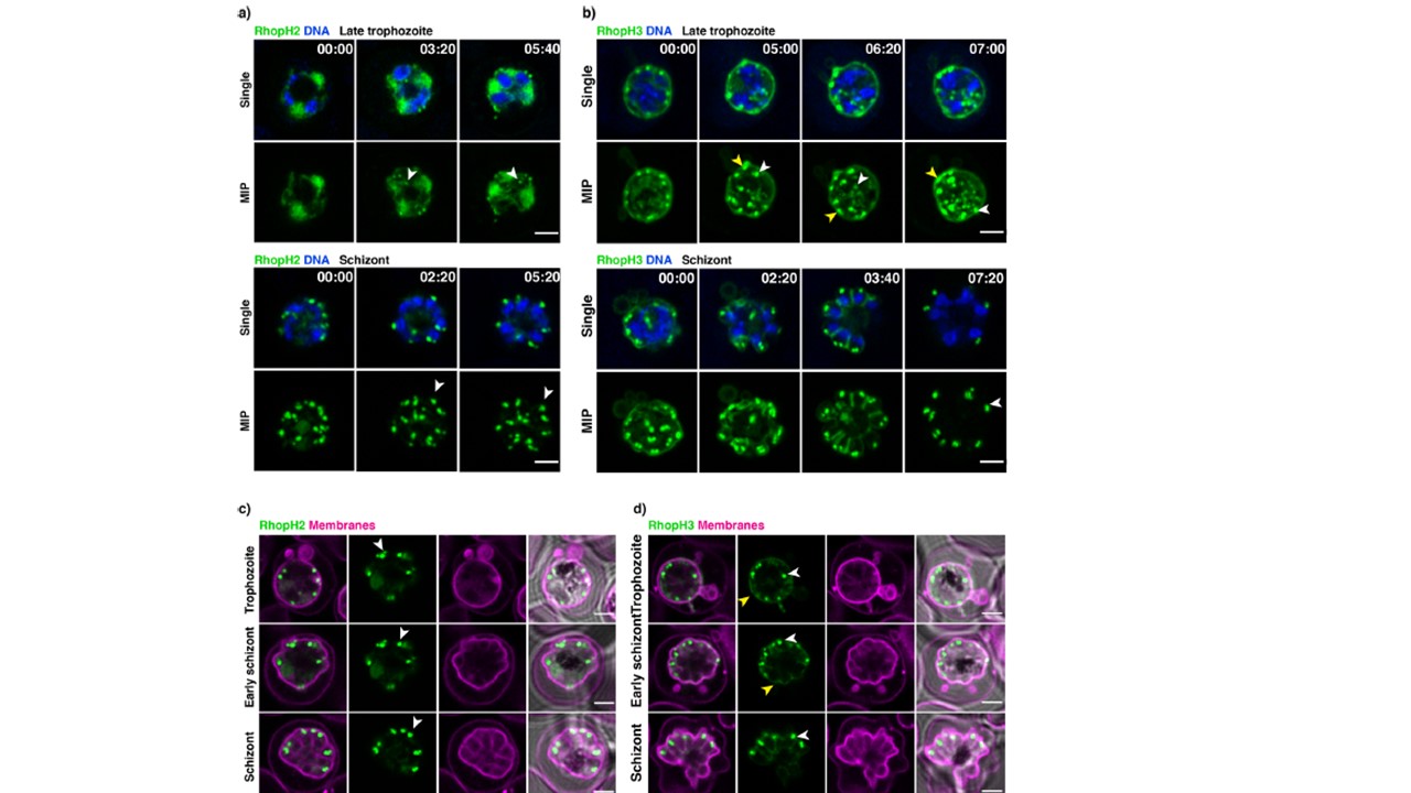
Super-resolution live imaging of RhopH2 and RhopH3. a Live RhopH2-mNeonGreen expressing trophozoites and schizont parasites with SiR-DNA stained nuclei (blue). mNeonGreen signal accumulation in forming rhoptries - white arrows. b Live RhopH3-mNeonGreen expressing trophozoites and schizont parasites with SiR-DNA-stained nuclei (blue). mNeonGreen signal accumulation in forming rhoptries (white arrows) and membrane association (yellow arrows). c Live RhopH2-mNeonGreen expressing trophozoites and schizont parasites with membrane dye (purple). d Live RhopH3-mNeonGreen expressing trophozoites and schizont parasites with membrane dye (purple). Each panel is a single z section; MIP – maximum intensity projections with a scale bar 2 μm. Time stamps in the upper-right corner represent time points of the overnight time-lapse experiment in the hours: minutes format.
Pasternak M, Verhoef JMJ, Wong W, Triglia T, Mlodzianoski MJ, Geoghegan N, Evelyn C, Wardak AZ, Rogers K, Cowman AF. RhopH2 and RhopH3 export enables assembly of the RhopH complex on P. falciparum-infected erythrocyte membranes. Commun Biol. 2022 5(1):333. PMID: 35393572.
Other associated proteins
| PFID | Formal Annotation |
|---|---|
| PF3D7_0929400 | high molecular weight rhoptry protein 2 |