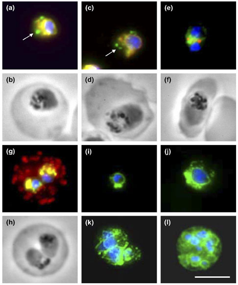
PfPDI localization in different parasite erythrocytic stages and co-localization with markers of subcellular compartments. (a,b) Immunostaining with antisera directed against rPfPDI (green) and PfBIP-PFI0875w (red) and the corresponding phase contrast image of a trophozoite. (c,d) Immunostaining with antisera directed against rPfPDI (green) and PfERC-PF11_0098 (red) and the corresponding phase contrast image of a trophozoite. (e,f) Immunostaining with antisera directed against rPfPDI (green) and PfERD2-PF13_0280 (red) and the corresponding phase contrast image of a young schizont. (g,h) Immunostaining with antisera directed against rPfPDI (green) and Pf332-PF11_0507 (red) and the corresponding phase contrast image of an erythrocyte infected by two young trophozoites. Regions of total overlapping of both proteins appear a yellow-orange color, DNA staining by DAPI appears in blue color. Arrows indicate the absence of co-localization.
(i-l) Overlaid images of parasites immunostained with antiserum directed against rPfPDI (green) and incubated with the nuclear dye DAPI (blue). Parasite stages were: ring (i), old trophozoites (j), schizont (k) and mature schizont with individualized merozoites (l). Bar: 5 mm. PfPDI with endoplasmic reticulum-resident proteins, PfBIP and PfERC, but not with the Golgi marker PfERD2 or the Maurer’s cleft marker Pf332-PF11_0507.
Mouray E, Moutiez M, Girault S, Sergheraert C, Florent I, Grellier P. Biochemical properties and cellular localization of Plasmodium falciparum protein disulfide isomerase. Biochimie. 2007 89:337-46. Copyright Elsevier 2009.