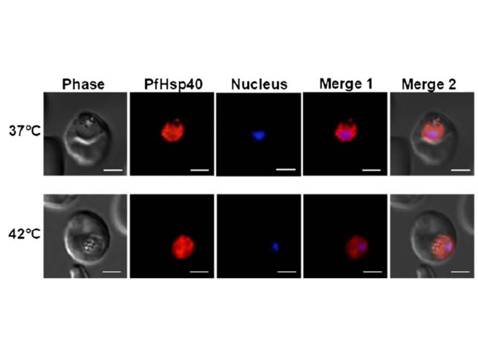
PfHsp40 resides mainly in the parasite cytosol. Immunofluorescence staining to detect PfHsp40 was conducted on trophozoite stage P. falciparum-infected erythrocytes. Upper panels parasite infected erythrocytes maintained at 37°C, lower panels parasite infected erythrocytes incubated at 42°C for 2 h prior to fixation and staining. PfHsp40 was detected using the rabbit anti-PfHsp40 antibody and Cy3-conjugated goat anti-rabbit secondary antibody (indicated in red). Parasite nuclei were stained with Hoechst (indicated in blue). Columns: Phase phase-contrast image, Nucleus parasite nuclei, PfHsp40 PfHsp40 localization, Merge 1 merged image indicating PfHsp40 localization relative to the parasite nucleus. The white size bars in each frame indicate 3 μm.
Botha M, Chiang AN, Needham PG, Stephens LL, Hoppe HC, Külzer S, Przyborski JM, Lingelbach K, Wipf P, Brodsky JL, Shonhai A, Blatch GL. Plasmodium falciparum encodes a single cytosolic type I Hsp40 that functionally interacts with Hsp70 and is upregulated by heat shock. Cell Stress Chaperones. 2011 16(4):389-401.