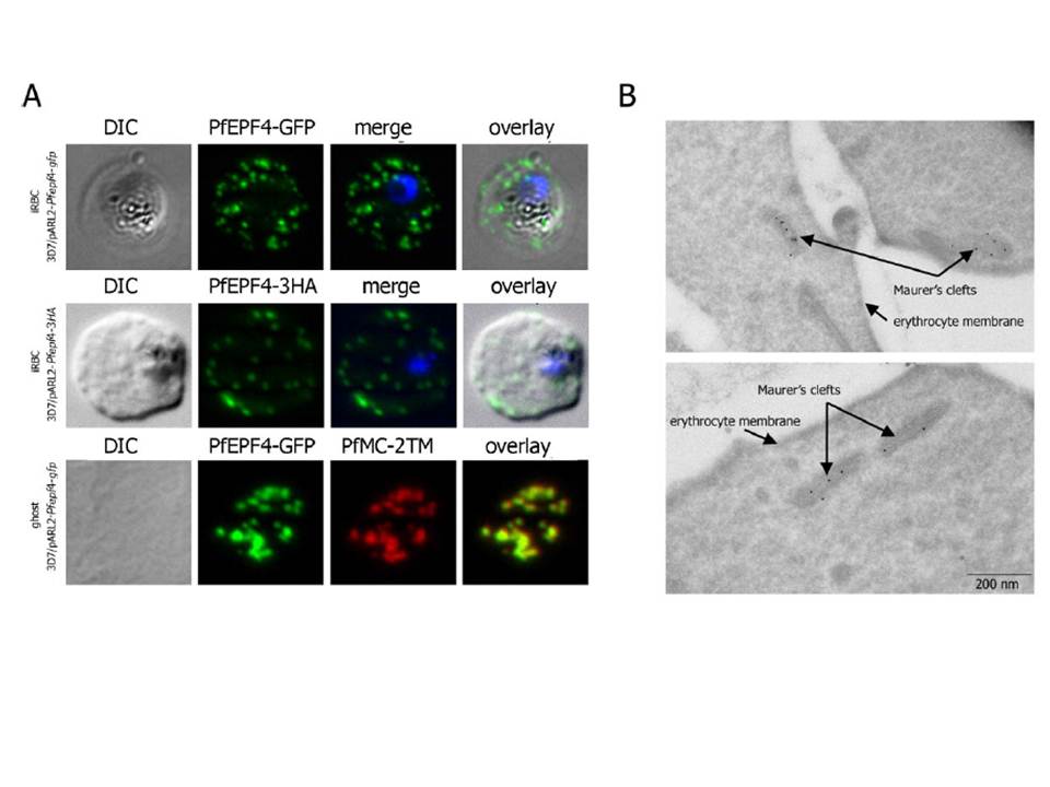
Localization and topology of the PfEPF4 proteins. A. Fluorescent patterns of iRBCs infected by 3D7/pARL2-Pfepf4-gfp (live imaging) and 3D7/pARL2-Pfepf4-3HA (immunodetection using anti-HA antibodies) and of resealed ghosts from 3D7/pARL2-Pfepf4-gfp iRBCs (GFP fluorescence, in green, and immunodetection using anti-PfMC-2TM antibodies, in red). RBCs and ghost preparations were incubated with DAPI for nucleus labelling. B. Immunoelectron microscopy using anti-GFP antibodies. PfEPF4 (A) display a pattern of fluorescent dots in the iRBC cytoplasm. PfEPF4-GFP colocalized with a Maurer’s cleft transmembrane protein, PfSBP1 or PfMC-2TM2 when resealed ghosts from iRBCs were analysed by immunofluorescence. The localization of PfEPF4 at the Maurer’s clefts was confirmed by immunoelectron-microscopy.
Mbengue A, Audiger N, Vialla E, Dubremetz JF, Braun-Breton C. Novel Plasmodium falciparum Maurer's clefts protein families implicated in the release of infectious merozoites. Mol Microbiol. 2013 88(2):425-42
Other associated proteins
| PFID | Formal Annotation |
|---|---|
| PF3D7_0101100 | exported protein family 4 Plasmodium exported protein (hyp5), unknown function |
| PF3D7_0631400 | Pfmc-2TM Maurer's cleft two transmembrane protein |