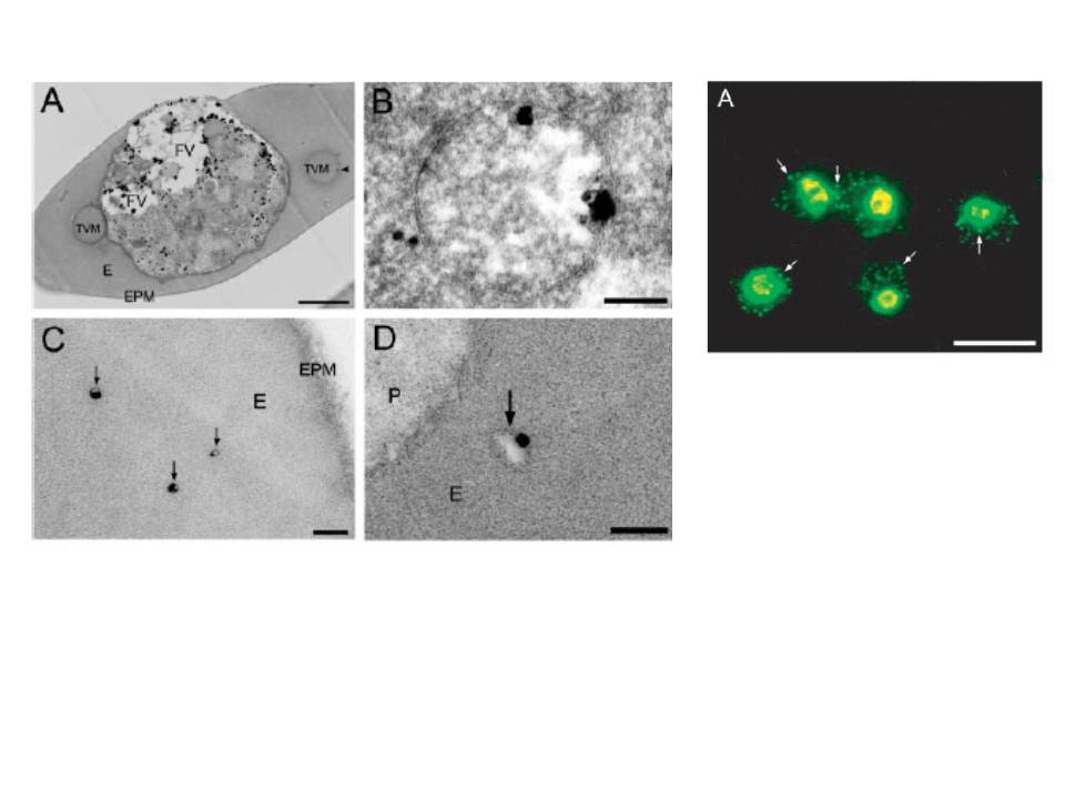
Left: Morphological evidence of the association of PfNSF with vesicular structures in parasitized erythocytes. Immunoelectromicroscopy for PfNSF was performed as described under “Experimental Procedures,” and the localization of PfNSF was visualized with immunogold particles after silver enhancement. The whole body of a parasitized erythrocyte (A), plastid-like organelle (B), or cytoplasm of erythrocyte (C and D) is shown. Vesicles containing PfNSF in erythrocyte cytoplasm are indicated by an arrowhead in A and by arrows in C and D. FV, parts of food vacuoles; E, erythrocyte cytoplasm; EPM, erythrocyte plasma membrane; P, parasite cytoplasm; TVM, tubovesicular membrane network. Bar, 1 mm in A and 0.2 mm in B–D.
Right: Parasitized erythrocytes were probed with anti-NSF Abs. Bar, 10 mm. Arrows indicate extra-parasitic vesicles.
Hayashi M, Taniguchi S, Ishizuka Y, Kim HS, Wataya Y, Yamamoto A, Moriyama Y. A homologue of N-ethylmaleimide-sensitive factor in the malaria parasite Plasmodium falciparum is exported and localized in vesicular structures in the cytoplasm of infected erythrocytes in the brefeldin A-sensitive pathway. J Biol Chem. 2001 276:15249-55.