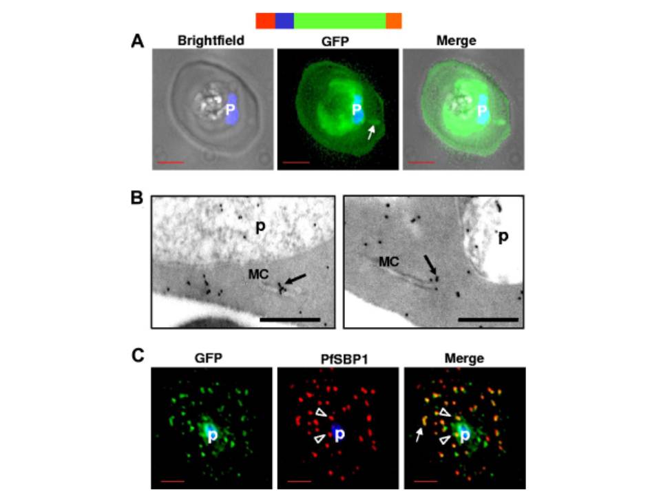
HTsol-GFP, a minimal soluble reporter exported to the erythrocyte cytoplasm and detected in clefts. (A) 0° projection of an erythrocyte infected with transgenic parasites expressing HTsol-GFP. Arrow indicates GFP-labeled intraerythrocytic structure, possibly a cleft. (B) Immunoelectron micrographs of trophozoite parasite (p)-infected cells expressing HTsol-GFP. Ultrathin sections were probed with antibodies to GFP and secondary antibody gold (10 nm) conjugate. Arrows indicate gold particles at intraerythrocytic Maurer’s clefts (MC). No gold labeling was detected in absence of primary antibody or when a nonspecific primary was used (not shown). Bar, 200 nm. (C) Single optical section of an infected erythrocyte expressing HTsol-GFP. Samples were treated to release soluble GFP, fixed, and probed with antibodies to GFP (green) and P falciparum Skeletal Binding Protein1 (PfSBP1, red). Arrow, GFP labeled cleft structures at the periphery of infected erythrocyte; arrowheads, clefts proximal to the parasite. In fluorescence micrographs, p denotes parasite nucleus stained with Hoechst 33342; bar, 2 mm. Schematic representation of the construct is indicated above with ER-type signal sequence (red), sequence containing HT signal (blue) fused to GFP (green) and myc (orange).
Bhattacharjee S, van Ooij C, Balu B, Adams JH, Haldar K. Maurer's clefts of Plasmodium falciparum are secretory organelles that concentrate virulence protein reporters for delivery to the host erythrocyte. Blood. 2008 111:2418-26.