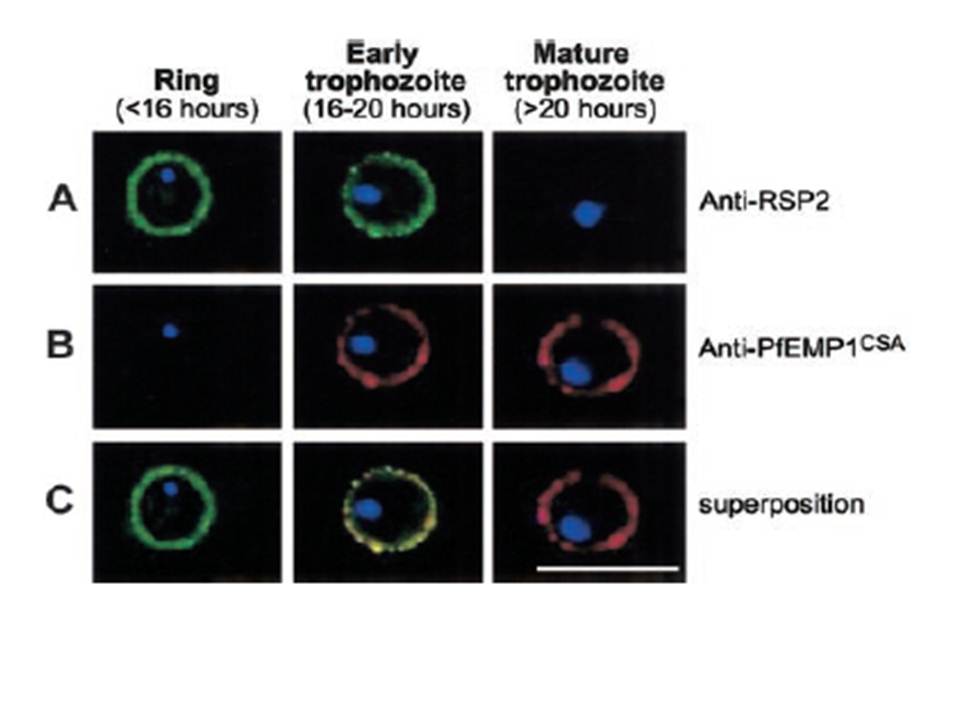
Expression profile of RSP2, the N terminal part of the Rap2 gene, and PfEMP1 at the erythrocyte surface during the blood-stage cycle. RSP-2CSA was stained with mAb B4 (green) and PfEMP1CSA with mAb 1B4/D4 (red) by L-IFA on IE. Parasite nuclei are stained by DAPI (blue). (A) Positive anti–RSP2 staining on synchronized ring stage–infected erythrocytes (16 hours after invasion) and early trophozoites (16 to 20 hours after invasion). No surface staining was detectable on mature forms (> 20 hours after invasion). (B) Absence of PfEMP1 staining on rings (16 hours) but strong IFA signal with early trophozoite and mature stages. (C) Superposition of anti–RSP2 and anti-PfEMP1 staining shows colocalization in early trophozoite stages. Scale bar measures 10 mm
Douki JB, Sterkers Y, Lépolard C, Traoré B, Costa FT, Scherf A, Gysin J. Adhesion of normal and Plasmodium falciparum ring-infected erythrocytes to endothelial cells and the placenta involves the rhoptry-derived ring surface protein-2. Blood. 2003 101:5025-32.
Other associated proteins
| PFID | Formal Annotation |
|---|---|
| PF3D7_1200600 | erythrocyte membrane protein 1, PfEMP1 |
