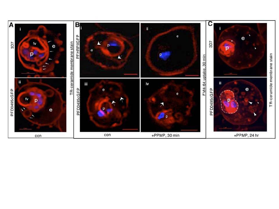
PFD0495c regulates the TVN in the infected erythrocyte cytoplasm. (A) Early schizonts (3D7 i and clone 1 parasites expressing PFD0495c-GFP ii) were stained with TR-ceramide (red; to visualize the TVN) and Hoechst 33342 (blue; to visualize DNA) and imaged live. Transgenic parasites display large TVN loops (arrows, panel ii) rather than tubules prominent in 3D7 (arrows, panel i). (B) Transgenic P. falciparum parasites expressing pfhrpii-gfp (i–ii) or pfd0495c-gfp (iii–iv) treated in absence (i, iii) or presence (ii, iv) of 5 mM PPMP for 30 min were incubated with membrane impermeable endocytic lipid marker FM4-64 (red) also for 30 min and imaged live. A second copy of pfd0495c promotes internalization of FM4-64 probe in the presence of PPMP (iv versus ii). (C) Ring stage parasites (3D7 i, pfd0495c-gfp clone 1 ii) were incubated with 5 mM PPMP for 24 hr, stained with TR-ceramide (red) and Hoechst (blue) and imaged live. Clone 1 accumulates an abundance of small loops and tubovesicular structures compared to few punctate spots in 3D7. Clone 1 parasites also show two nuclei (blue) and an almost two fold increase in size compared to 3D7 (that contain only one nucleus). Arrows show tubules, loops; arrowheads, vesicular structures; small arrowheads, punctate spots. For all panels erythrocyte (e), parasite (p) and intraerythrocytic structures (arrow). Scale bar, 2 mm.
Tamez PA, Bhattacharjee S, van Ooij C, Hiller NL, Llinás M, Balu B, Adams JH, Haldar K. An erythrocyte vesicle protein exported by the malaria parasite promotes tubovesicular lipid import from the host cell surface. PLoS Pathog. 2008 Aug 8;4(8):e1000118. PMID: