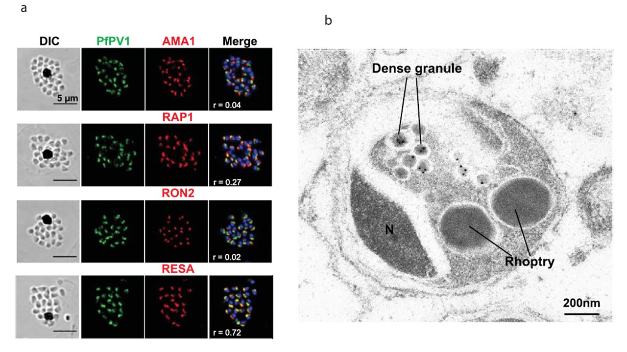
Subcellular localization of PfPV1 in schizont-stage parasites. (a) Results of IFA with Confocal Microscopy. Labeling of the utilized antibodies is on the upside of panels. PfPV1 (green) was counter stained with four different markers of subcellular localizations (red). The parasite nucleus was stained with DAPI (blue).
PfPV1 co-localized with RESA a dense granule marker. DIC; Differential interference contrast microscope. Merge; the image created by merging image of IFA, and nuclear-staining. Pearson’s coefficient values ( = r) were calculated by using Zen 2010 software (Carl Zeiss MicroImaging, Thornwood, NY) and shown on the merged panels. Scale bar = 5 μm. (b) Results of IEM using rabbit anti-PfPV1 antibody. Gold particles can be observed at the Plasmodium falciparum merozoite dense granules at schizont stage. N; nuclear. Scale bar = 200 nm.
Morita M, Nagaoka H, Ntege EH, Kanoi BN, Ito D, Nakata T, Lee JW, Tokunaga K, Iimura T, Torii M, Tsuboi T, Takashima E. PV1, a novel Plasmodium falciparum merozoite dense granule protein, interacts with exported protein in infected
erythrocytes. Sci Rep. 2018 Feb 27;8(1):3696
Other associated proteins
| PFID | Formal Annotation |
|---|---|
| PF3D7_0102200 | ring-infected erythrocyte surface antigen |
| PF3D7_1133400 | apical membrane antigen 1 |
| PF3D7_1410400 | rhoptry-associated protein 1 |
| PF3D7_1452000 | rhoptry neck protein 2 |