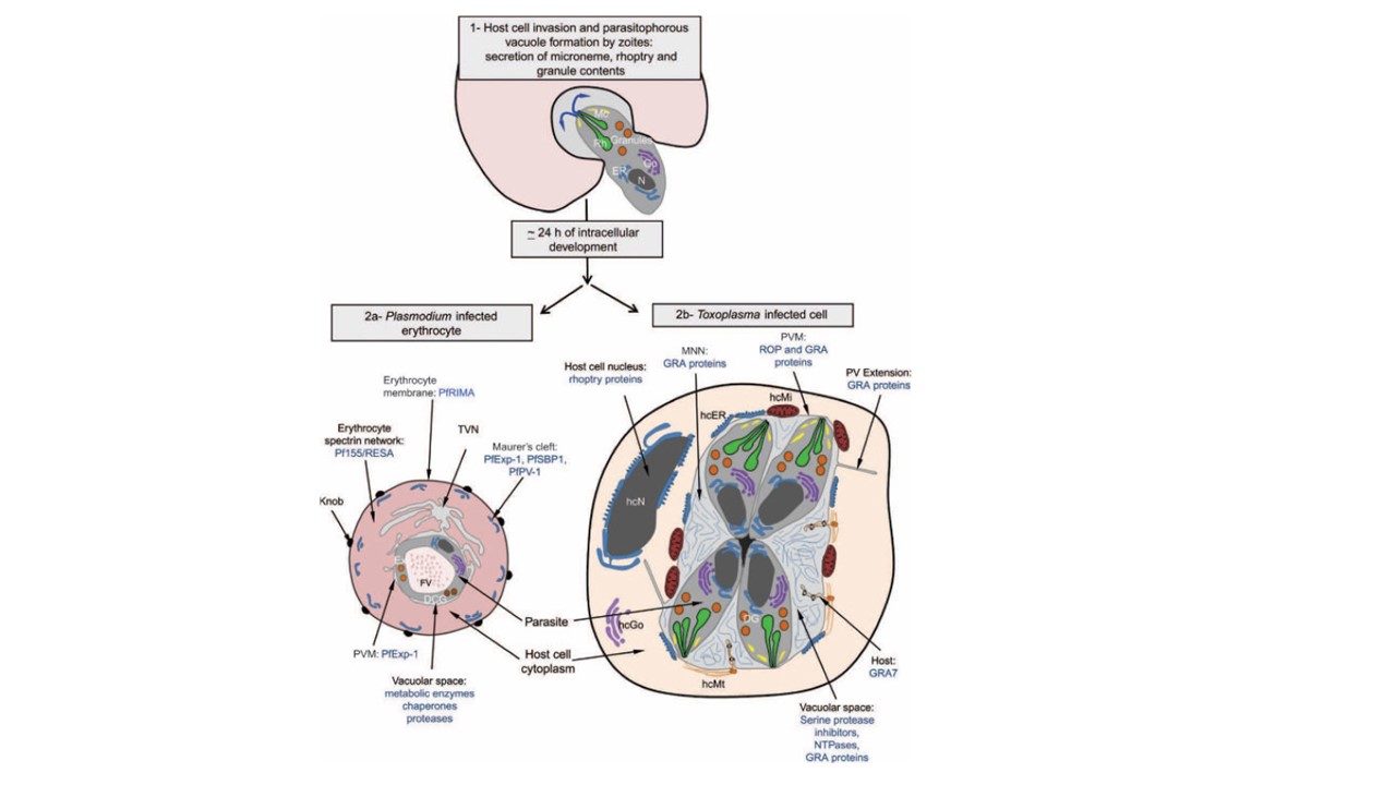
Protein delivery into the PV and the infected host cell of Toxoplasma gondii and Plasmodium falciparum. The entry mechanism and initial formation of the PV rely on machineries that are conserved across the phylum (1). In contrast, the diversity of dense core granule populations among Apicomplexa parasites likely reflects an evolutionary divergence required for specialized intracellular development. The Plasmodium PV is limited by the PVM, which is prolonged by large membranous extensions known as TVN into the erythrocyte’s cytosol. The MCs that are flattened structures within the host cell cytosol would constitute a transitory step for many proteins en route to the erythrocyte’s surface. Examples of the proteins identified in each compartment are provided (2a). The Toxoplasma PV is limited by the PVM. The intravacuolar space is decorated with the MNN. PVM extensions into the host cell as well as invaginations of the PVM known as HOSTs are also shown. The HOSTs contain a single host cell microtubule (hcMt). Proteins exported from both the rhoptries (Rh) and the dense granules (DG) were identified as being targeted to the PV space, the MNN, the HOSTs, the PVM and its extensions into the host cell as well as the host cell nucleus (hcN). Examples of these proteins are listed at their final destination (2b). DCG, dense core granules; ER, parasite endoplasmic reticulum; Ex, exonemes; FV, food vacuole; Go, parasite Golgi apparatus; hcER, host cell endoplasmic reticulum; hcGo, host cell Golgi apparatus; Mc, micronemes; N, parasite nucleus. Cesbron-Delauw MF, Gendrin C, Travier L, Ruffiot P, Mercier C. Apicomplexa in mammalian cells: trafficking to the parasitophorous vacuole. Traffic. 2008 9(5):657-64. PMID: 18315533.
