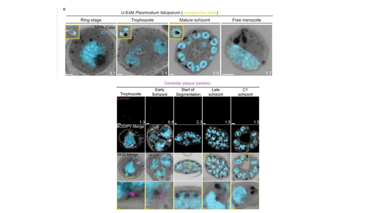
Characterisation of intranuclear and subpellicular microtubules.
3D7 parasites were prepared by U-ExM, stained with NHS ester (greyscale), BODIPY TRc (white), SYTOX (cyan) and anti-tubulin (microtubules; magenta) antibodies, and imaged using Airyscan microscopy. (a) Images of whole parasites throughout asexual blood-stage development. (b) Nuclei in the process of dividing, with their centriolar plaques connected by an interpolar spindle. (c) The number and type of microtubule branches in interpolar spindles and (d) length of interpolar microtubules. (e) Subpellicular microtubules (SPMTs) stained with an anti-poly-glutamylation (PolyE; yellow) antibody. (f) Quantification of the number of SPMTs per merozoite from C1-treated schizonts. (g) SPMT biogenesis throughout segmentation. (h) Model for SPMT biogenesis. PPM = parasite plasma membrane, APRs = apical polar rings, BC = basal complex, CP = centriolar plaque. Images are maximum-intensity projections, number on image = Z-axis thickness of projection in μm. Scale bars = 2 μm. Liffner B, Cepeda Diaz AK, Blauwkamp J, Anaguano D, Frolich S, Muralidharan V, Wilson DW, Dvorin JD, Absalon S. Atlas of Plasmodium falciparum intraerythrocytic development using expansion microscopy. Elife. 2023 12:RP88088. PMID: 38108809;