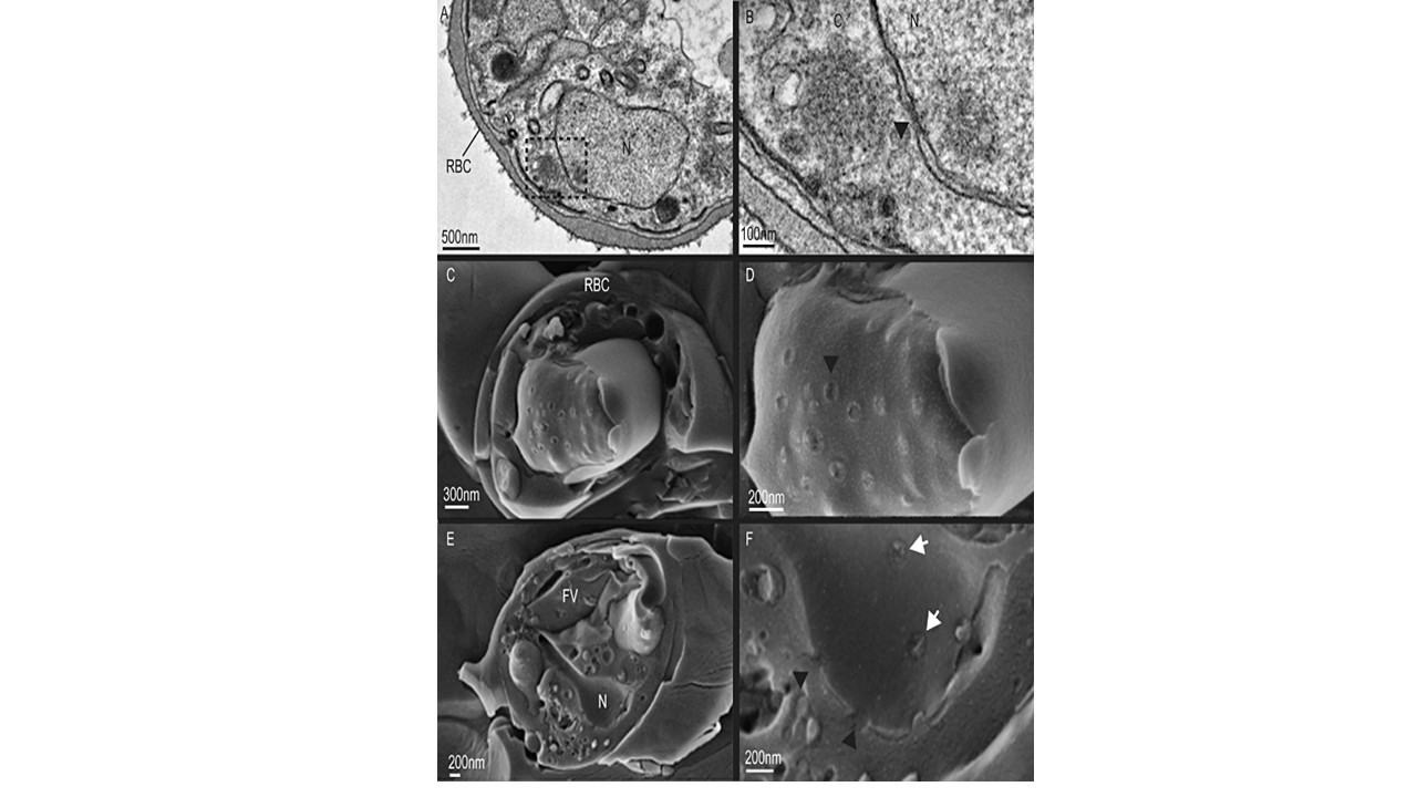
A and B. Cross-section through a red blood cell infected with an early schizont obtained by TEM of chemically fixed iRBC. The framed region in (A) is presented at higher magnification in (B) and shows a nuclear pore with its inner ring seen as electron-dense matter between the double membranes of the nuclear envelope.
C and D. Freeze fracture cryoSEM exposing the outer surface of an early schizont nucleus with a cluster of NPCs (arrowhead). E and F. Freeze fracture cryoSEM through an early schizont exposing the inner part of the nuclear membrane. The framed region in (E) is presented at higher magnification in F and shows two NPCs (arrowed). Two more NPCs were exposed in a longitude break where the surrounding nuclear envelope can be observed (arrowhead). Weiner A, Dahan-Pasternak N, Shimoni E, Shinder V, von Huth P, Elbaum M, Dzikowski R. 3D nuclear architecture reveals coupled cell cycle dynamics of chromatin and nuclear pores in the malaria parasite Plasmodium falciparum. Cell Microbiol. 2011 13(7):967-77.
c
