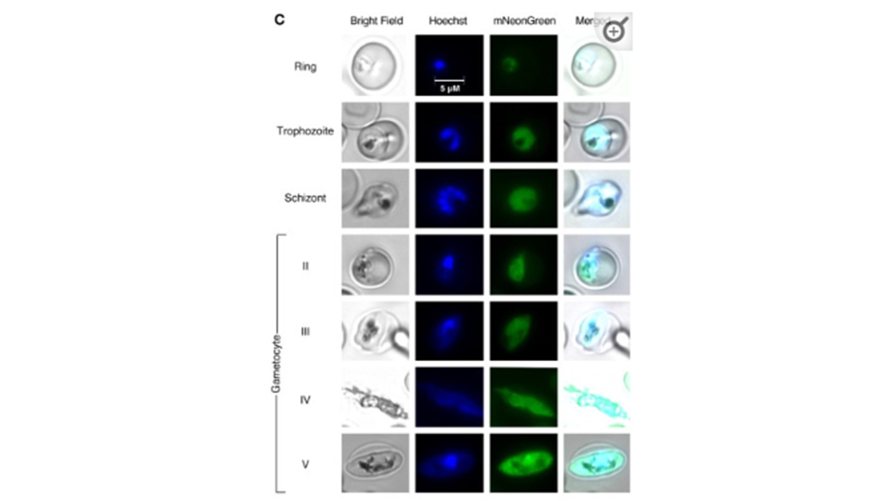
Fluorescence microscopy of synchronised mNeonGreen@pare parasites at asexual ring, trophozoite and schizont stages, and at gametocyte stages II-V (obtained through gametocytogenesis induction). Cultures were fixed, stained with Hoechst DNA stain, and visualised using bright-field and fluorescence microscopy at 1000x magnification.
Hoshizaki J, Jagoe H, Lee MCS. Efficient generation of mNeonGreen Plasmodium falciparum reporter lines enables quantitative fitness analysis. Front Cell Infect Microbiol. 2022 12:981432. PMID: 36189342