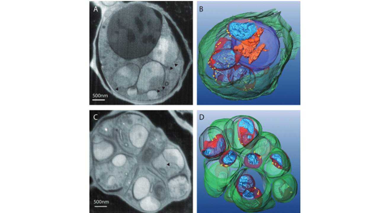
3D analysis of P. falciaprum nuclei in mid and late schizonts by serial surface view imaging shows reorientation of nuclear pores (FIB‘ slice and view’). Contrast is inverted with respect to TEM, so that the dense genetic matter is brighter and the loose genetic matter is relatively dark. A. A slice through a parasite in mid schizont stage. NPCs are also seen here (arrowheads).B. 3D model of the entire parasite in mid schizont stage. Red – nuclear membrane; yellow – NPC; blue – heterochromatin; purple – food vacuole; orange – haemozoin; green – RBC membrane. NPCs are oriented outward, facing the parasite membrane. C. A slice through a parasite in late schizont stage. NPC is also seen here (arrowhead).D. 3D model of the entire parasite in late schizont stage; red – nuclear membrane; yellow – NPC; blue – heterochromatin; green – merozoite membrane. NPCs are oriented to face the cytoplasm of each incipient merozoite. 3D nuclear architecture reveals coupled cell cycle dynamics of chromatin and nuclear pores in the malaria parasite Plasmodium falciparum.
Weiner A, Dahan-Pasternak N, Shimoni E, Shinder V, von Huth P, Elbaum M, Dzikowski R. 3D nuclear architecture reveals coupled cell cycle dynamics of chromatin and nuclear pores in the malaria parasite Plasmodium falciparum. Cell Microbiol. 2011. PMID: 21501361.