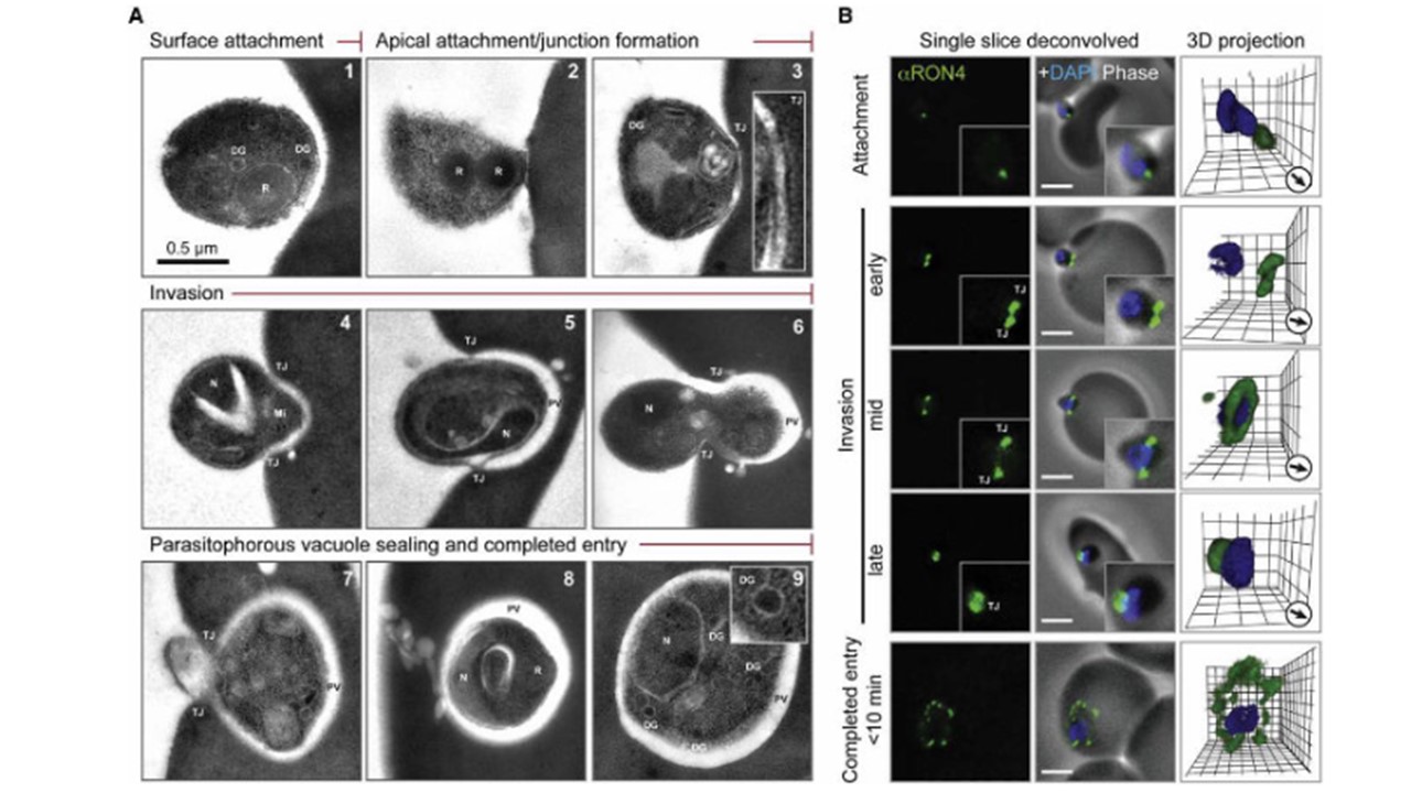
Time courses of invasion by transmission electron microscopy (TEM) (A) and wide-field immunofluoresence assay (IFA) microscopy (B) with deconvolution (single slice or three-dimensional reconstruction) labeled with PfRON4. For TEM numbering, see the main text. The IFA scale bar represents 2.0 mm. In 3D images, gamma settings were altered, and the grid represents 0.5 mm intervals. DG, dense granules; Mi, micronemes; N, nucleus; PV, parasitophorous vacuole; R, rhoptries; TJ, tight junction. Riglar DT, Richard D, Wilson DW, Boyle MJ, Dekiwadia C, Turnbull L, Angrisano F, Marapana DS, Rogers KL, Whitchurch CB, Beeson JG, Cowman AF, Ralph SA, Baum J. Super-resolution dissection of coordinated events during malaria parasite invasion of the human erythrocyte. Cell Host Microbe. 2011 PMID: 21238943