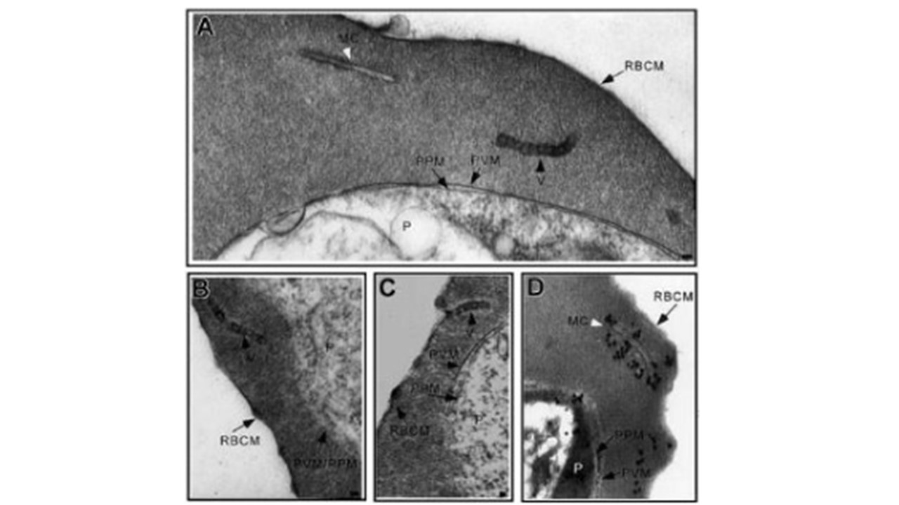
Incubation of IRBCs with AlF reveals vesicle chains. (A-B) Ultrastruc-
tural characterization of glutaraldehyde-fixed trophozoite stage IRBCs revealed vesicles with an electron-dense limiting membrane arranged in chains in the RBC cytosol (black arrowhead) or (C) closely apposed to the erythrocyte plasma membrane. A typical Maurer cleft is observed (A; white arrowhead). (D) Immunoelectron microscopy confirmed the long, slender unit membranes in the RBC cytosol are Maurer clefts. MC indicates Maurer cleft; P, parasite; PVM, parasitophorous vacuolar membrane; PPM, parasite plasma membrane, RBCM, red blood cell membrane; and V, vesicles. Scale bar = 70 nm. Taraschi TF, O'Donnell M, Martinez S, Schneider T, Trelka D, Fowler VM, Tilley L, Moriyama Y. Generation of an erythrocyte vesicle transport system by Plasmodium falciparum malaria parasites. Blood. 2003 PMID: 12869498.