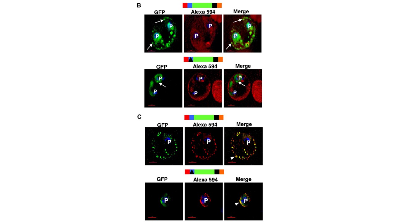
The HT motif sorts protein into Maurer’s clefts without translocation across the PVM. (B) Single optical sections of ghosts resealed with Alexa Fluor 594 anti-GFP antibodies infected with parasites expressing HT-GFPmembmyc (top panel) or D-GFPmembmyc (bottom panel). Cells were viewed live using optics for GFP (green), Alexa Fluor 594/Rhodamine (red), and the merged image is shown in the right panel. Arrows, GFP labeled clefts not labeled with anti-GFP Alexa Fluor 594 conjugate. (C) Immunofluorescence assay of resealed ghosts infected with parasites expressing HT-GFPmembmyc (top panel) or D-GFPmembmyc (bottom panel) permeabilized with saponin and treated with anti-GFP Alexa Fluor 594-conjugated antibodies. Images under GFP (green) and Alexa 594 (red) optics and their respective merge are shown. Arrowhead, region of colocalization (in yellow) between GFP and Alexa 594. In all cells, the parasite (p) nuclei were stained with Hoechst 33342 (blue); bar, 2 mm. Schematic representation of the construct is indicated above with ER-type signal sequence (red), sequence containing HT signal (blue) or its replacement (filled triangle in black) fused to GFP (green), transmembrane region (black), and myc (orange Bhattacharjee S, van Ooij C, Balu B, Adams JH, Haldar K. Maurer's clefts of Plasmodium falciparum are secretory organelles that concentrate virulence protein reporters for delivery to the host erythrocyte. Blood. 2008 111(4):2418-26. PMID: 18057226;