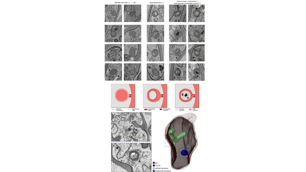
A Exemplar micrographs of the cytostome in 20 different gametocytes subdivided into three categories. Left: Cytostome without further membrane delineation in lumen, relatively homogenous lumen and circular grooves that could indicate future emergence of ring. Middle: Relatively large membrane electron dense ring and small electron lucent lumen. Ring is continuous with RBC cytosol, putative intermediate state. Right: Electron dense ring with homogenous thickness and lumen with heterogeneous content. Ring is continuous with RBC cytosol. All categories have close association of ER unlike the ABS cytostome. ER = light green square, halo/membrane ring = red arrow, yellow asterisk = separate lumen, plus sign = osmiophilic content, scale bars = 0.1 μm. B Schematic of the hypothesized cytostome variants/intermediates. Each schematic corresponds to the variant above. C Rendering of a developing gametocyte that contains both a canonical ABS cytostome and a cytostome with distinct ring and lumen. The ABS cytostome (green) is characterized by homogenous dark lumen and continuation of the parasite membrane that forms the invagination. The gametocyte cytostome (blue) is characterized by a bulbous shape and distinct electron dense ring with electron lucent lumen. Coexistence of ABS-like and gametocyte cytostome appears uncommon and has been observed in three cells throughout all stacks. Scale bars = 0.5 μm. Evers F, Roverts R, Boshoven C, Kea-Te Lindert M, Verhoef JMJ, Sommerdijk N, Sinden RE, Akiva A, Kooij TWA. Comparative 3D ultrastructure of Plasmodium falciparum gametocytes. Nat Commun. PMID: 39747010