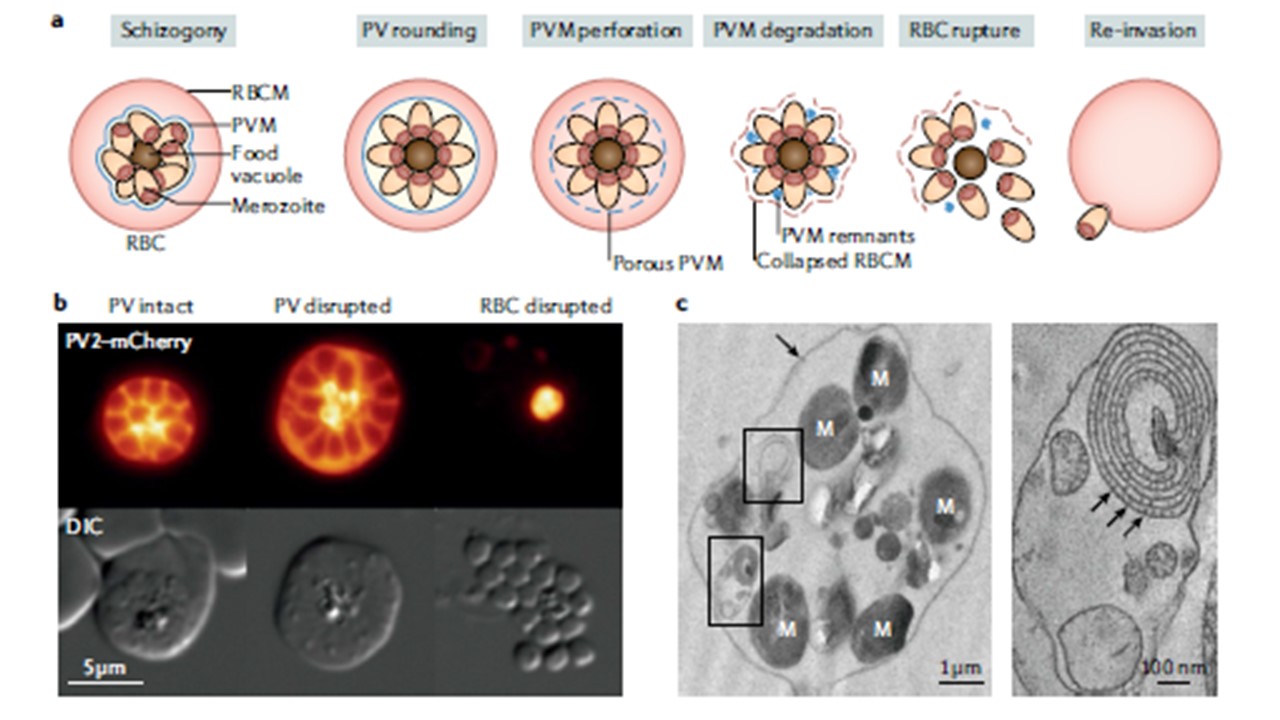
The parasitophorous vacuole (PV) membrane (PVM) (blue) undergoes distinct morphological phases during parasite egress. On completion of schizogony , the merozoites align around the central food vacuole (dark brown) and the parasitophorous vacuole rounds up. The PVM becomes perforated and is then broken down into multilamellar vesicles, which coincides with red blood cell (RBC) membrane (RBCM) poration and collapse. Subsequent RBCM rupture releases the merozoites and the cycle starts anew. b | Equilibration of soluble protein contents across the PVM before merozoite exit. Shown are live fluorescence micrographs of transgenic Plasmodium berghei schizonts expressing mCherry-tagged PV2 (orange) before (left), during (middle) and after (right) parasite egress. Initially , fluorescence is closely associated with the merozoite periphery , then spreads throughout the RBC cytoplasm on PVM disruption and ultimately diffuses into the extracellular medium, leaving only a brightly stained fraction within the parasite’s digestive vacuole on completed egress. c | Fragments of ruptured PVM (black boxes) are observed within the RBCM (black arrow) of E64-treated Plasmodium falciparum schizonts by transmission electron microscopy of high-pressure frozen, freeze-substituted thin sections (left). PVM fragments frequently form multilamellar vesicles (right), within which PVM-associated protein complexes can be observed (black arrows). Shown is an average of ten central slices from a tomogram reconstructed from a −60° to +60° dual-axis tilt series collected with a 120-kV electron microscope. DIC, differential interference contrast; M, merozoite. Electron microscopy images courtesy of Claudine Bisson, Birkbeck College, University of London, UK. Matz JM, Beck JR, Blackman MJ. The parasitophorous vacuole of the blood-stage malaria parasite. Nat Rev Microbiol. 2020. PMID: 31980807
PubMed Article: The parasitophorous vacuole of the blood-stage malaria parasite
