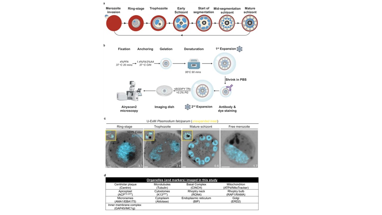
(U-ExM) workflow and summary of parasite structures imaged in this study. (a) Diagram of asexual blood-stage lifecycle of P. falciparum. (b) U-ExM workflow used in this study. PFA = paraformaldehyde, FA = formaldehyde, AA = acrylamide, PG = propyl gallate. Snowflake indicates steps where gels were cryopreserved. (c) Comparison of brightfield and DAPI staining of unexpanded P. falciparum parasites (inset) with P. falciparum prepared by U-ExM, stained with N-hydroxysuccinimide (NHS) ester (protein density; grayscale) and SYTOX Deep Red. (DNA; cyan), and imaged using Airyscan microscopy. Images are maximum-intensity projections, number on image = Z-axis thickness of projection in μm. Scale bars = 2 μm. (d) Summary of all organelles, and their corresponding antibodies, imaged by U-ExM in this study. Liffner B, Cepeda Diaz AK, Blauwkamp J, Anaguano D, Frölich S, Muralidharan V, Wilson DW, Dvorin J, Absalon S. Atlas of Plasmodium falciparum intraerythrocytic development using expansion microscopy. 2023