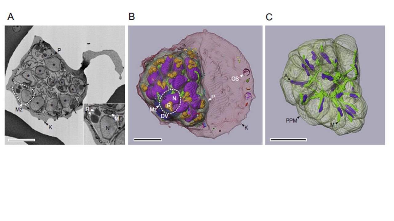
A SBF-SEM image and the 3D reconstruction of a late schizont stage-iRBC. (A) A SBF-SEM image. Inset in (A) shows one of merozoites formed within the schizont-stage parasite. (B) A whole 3D image. (C) The 3D reconstruction of a parasite plasma membrane (PPM), a mitochondrion (M), and apicoplasts (A). The SBF-SEM map has been deposited under accession number EMPIAR-10055. P, parasite; N and asterisks, nucleus; Mz, merozoite; R and black arrows, rhoptry; DV, digestive vacuole; OS, oval membranous structure; K, knob. All scale bars = 2 mm.
Sakaguchi M, Miyazaki N, Fujioka H, Kaneko O, Murata K. Three-dimensional analysis of morphological changes in the malaria parasite infected red blood cell by serial block-face scanning electron microscopy. J Struct Biol. 2016 Mar;193(3):162-71. PMID: 26772147.
