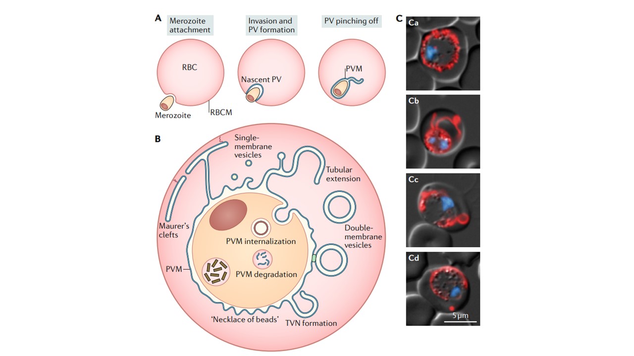
A | Biogenesis of the parasitophorous vacuole (PV). The merozoite induces invagination of the red blood cell membrane (RBCM) during invasion. Active merozoite motility and subsequent membrane scission result in the formation of the parasitophorous vacuole membrane (PVM) (blue). b | Remodelling and trafficking of the PVM. The vacuole can form a continuous space around the parasite. The frequently observed ‘necklace of beads’ morphology is indicative of constriction zones between the parasite and the PVM. Membrane whorls emerge from the PVM and envelop host cell cytoplasm, resulting in formation of the tubovesicular network (TVN) and the release of double-membrane vesicles. The TVN also features distinct junctional sites (green), as well as partially interconnected tubular and vesicular compartments. Maurer’s clefts are believed to bud off from the parasitophorous vacuole and become tethered below the RBCM through proteinaceous anchors (red lines extending from the RBCM). There is debate as to whether the Maurer’s clefts remain connected to the vacuole. Endocytosis of host cell cytoplasm leads to the formation of intraparasitic endosomes lined by the PVM and parasite plasma membrane (PPM). The PVM is ultimately degraded during haemoglobin digestion and concomitant haemozoin formation (brown rectangles). c| Morphology of the parasitophorous vacuole in maturing parasites. Shown are live fluorescence micrographs of transgenic Plasmodium berghei parasites expressing mCherry-tagged PV1 (parts ca and cb) or EXP2 (parts cc and cd), showing the ‘necklace of beads’ (part ca), tubular extensions (part cb), TVN whorls (part cc) and budding from the PVM (part cd). Red indicates tagged protein and blue indicates the nucleus.
Matz JM, Beck JR, Blackman MJ. The parasitophorous vacuole of the blood-stage malaria parasite. Nat Rev Microbiol. 2020 Jan 24. PMID: 31980807.