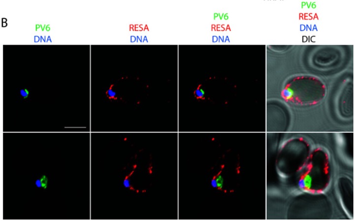
Role of Plasmepsin V in the processing of PV6 and localization of PV6 after invasion. (B) Localization of PV6 and RESA immediately after invasion of 3D7 parasites. Invasion was synchronized with ML10, and samples were collected within 2 h after the removal of ML10. Note that the erythrocyte at the top of the panel is also infected and therefore also displays anti-RESA staining but that the parasite in that cell is outside of the plane of focus. The scale bar represents 5 µm.
Fréville A, Ressurreição M, van Ooij C. Identification of a non-exported Plasmepsin V substrate that functions in the parasitophorous vacuole of malaria parasites. mBio. 2024 Jan 16;15(1):e0122323.
PubMed Article: Identification of a non-exported Plasmepsin V substrate that functions in the parasitophorous vacuole of malaria parasites
Other associated proteins
| PFID | Formal Annotation |
|---|---|
| PF3D7_0102200 | ring-infected erythrocyte surface antigen |
