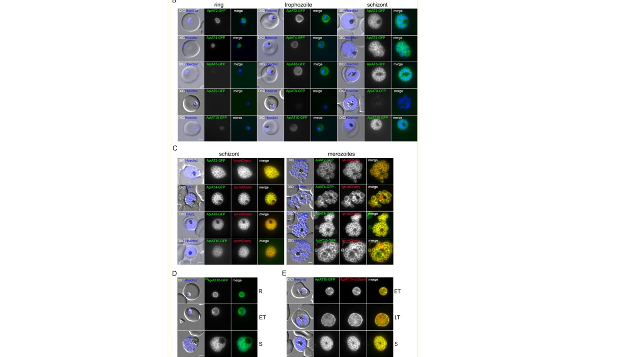
P. falciparum ApiATs localize to the parasite plasma membrane (PPM) during asexual blood stage development. (B) Localization of PfApiAT2-GFP, PfApiAT4-GFP, PfApiAT8-GFP, PfApiAT9-GFP, and PfApiAT10-GFP by live-cell microscopy across the IDC of 3D7 parasites. DIC, differential interference contrast. (C) Colocalization of the GFP-tagged ApiAT fusion proteins with the PPM marker protein Lyn-mCherry in schizonts and merozoites. (D) Live-cell microscopy of 3D7-crt-ApiAT10-GFP parasites across the IDC. (E) 3D7-iGP-ApiAT10-GFP/ama1-ApiAT10-mCherry parasites at the trophozoite and schizont stage. Nuclei were stained with Hoechst-33342. Parasite stages are indicated as follows; ring stage (R), early trophozoite (ET), late trophozoite (LT) and schizont (S). Bars, 2 μm. Wichers JS, van Gelder C, Fuchs G, Ruge JM, Pietsch E, Ferreira JL, Safavi S, von Thien H, Burda PC, Mesén-Ramirez P, Spielmann T, Strauss J, Gilberger TW, Bachmann A. Characterization of Apicomplexan Amino Acid Transporters (ApiATs) in the Malaria Parasite Plasmodium falciparum. mSphere. 2021 6(6):e0074321. PMID: 34756057;
Other associated proteins
| PFID | Formal Annotation |
|---|---|
| PF3D7_0104700 | apicomplexan amino acid transporter apiat9, putative |
| PF3D7_0312500 | apicomplexan amino acid transporter apiat10 |
| PF3D7_0914700 | apicomplexan amino acid transporter apiat2, putative |
| PF3D7_1129900 | apicomplexan amino acid transporter apiat4 |
