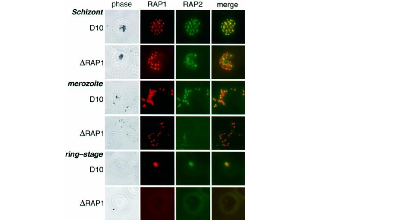
RAP1 specifies rhoptry localization of RAP2. Individual rows show D10 and D10ΔRAP1c1 schizonts, merozoites and ring-stage parasites, as labelled. Each row shows phase-contrast images, staining with polyclonal rabbit anti-RAP1 (red), staining with monoclonal anti-RAP2 (green), and merged RAP1 + RAP2 staining (yellow indicates co-localization) for the same parasite. Top panel: D10 schizont showing punctate rhoptry localization of both RAP1 and RAP2. Second panel: the truncated RAP1 protein in D10ΔRAP1c1 schizonts (see Figure 2) still localizes to the rhoptries, but RAP2 exhibits diffuse non-rhoptry fluorescence. Third panel: RAP1 and RAP2 co-localize in D10 merozoites. Fourth panel: RAP1 exhibits similar (presumably rhoptry) focal staining in D10ΔRAP1c1 parasites, but RAP2 is not detectable. Fifth panel: in wild-type parasites, both RAP1 and RAP2 proteins are carried into ring-stage parasites after infection. Sixth panel: neither RAP1 nor RAP2 are carried into the erythrocyte during merozoite infection. Ring-stage parasites were identified by staining with DAPI (not shown). Baldi DL, Andrews KT, Waller RF, Roos DS, Howard RF, Crabb BS, Cowman AF. RAP1 controls rhoptry targeting of RAP2 in the malaria parasite Plasmodium falciparum. EMBO J. 2000 19(11):2435-43. PMID: 10835342
Other associated proteins
| PFID | Formal Annotation |
|---|---|
| PF3D7_0501600 | rhoptry-associated protein 2 |