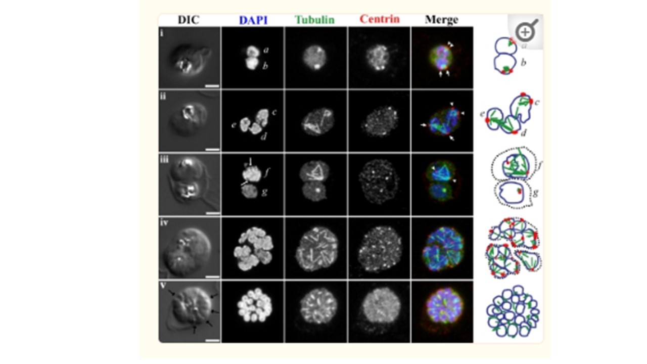
Fluorescence light microscopy images of mitotic spindle during blood-stage schizogony in P. falciparum parasites. 3D confocal microscopy views of blood-stage P. falciparum parasite cell morphology (DIC microscopy), nuclei (DAPI, blue in merged images), mitotic spindle (anti-alpha-tubulin antibody, green in merged images), and mitotic spindle MTOCs (anti-PfCEN3 antibody in rows i, ii, iv, and v or anti-Cr Centrin1 antibody 20H5 in row iii, red in merged images) as previously described (42). Schematic cartoons are drawn for each example, indicating spindle MTOCs (red circles), microtubules (green lines), and outlines of stained DNA (blue lines). Dotted black lines indicate separate parasites in multiply infected host cells. Row i, a and b, short mitotic spindles bounded by MTOCs. The spindle in nucleus b which resembles an oblong tubulin spot has a pole-to-pole distance that is approximately 1 μm long, similar to the lengths of anaphase spindles as described by TEM. Rows ii and iii, c to f, larger spindle structures. In d and e, spindle extends across separate nuclear bodies. In f, a dark line is visible down the center of the DAPI-stained DNA near the spindle midzone (DAPI, arrows). Row iv, asynchronous schizont nuclei have spindles with different geometries in multiple stages of extension. Row v, segmented parasites after the final mitosis of schizogony. Furrows are visible between daughters (arrows), nuclei are condensed, and microtubules appear in the daughter cytoplasms. Row i is a single confocal slice from a 3D series. Rows ii to v are maximum-projection images of full 3D series. Rows ii to iv were processed with deconvolution software. Scale bar, 2 μm.
Gerald N, Mahajan B, Kumar S. Mitosis in the human malaria parasite Plasmodium falciparum. Eukaryot Cell. 2011 10(4):474-82. PMID: 21317311
Other associated proteins
| PFID | Formal Annotation |
|---|---|
| PF3D7_1475700 | tubulin epsilon chain, putative |