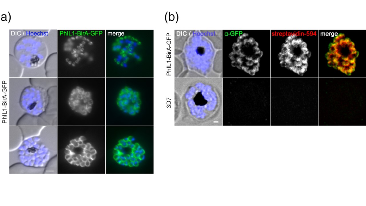
Identification of PhIL1-interacting candidates using BioID. (a) IMC localisation of PfPhIL1-BirA-GFP in schizonts and merozoites. Nuclei were stained with Hoechst-33,342. Scale bar, 2 μm. Widefield fluorescence microscopy confirmed the expected IMC localization pattern of PfPhIL1-BirA-GFP with an additional weak expression in ring stages(b) PfPhIL1-based biotinylation at the IMC. PfPhIL1-BirA-GFP (mouse anti-GFP, green) or 3D7 control parasites were fixed with MeOH at the schizont stage and biotinylated proteins were visualized by streptavidin-594 (red). We visualized biotinylated proteins with fluorescent-labelled streptavidin by fluorescent microscopy revealing colocalization with our IMC marker PfPhiL1-BirA-GFP with no background staining in 3D7 control parasites. Hoechst was used for DNA staining (blue). Scale bar, 1 μm.
Wichers JS, Wunderlich J, Heincke D, Pazicky S, Strauss J, Schmitt M, Kimmel J, Wilcke L, Scharf S, von Thien H, Burda PC, Spielmann T, Löw C, Filarsky M, Bachmann A, Gilberger TW. Identification of novel inner membrane complex and apical annuli proteins of the malaria parasite Plasmodium falciparum. Cell Microbiol. 2021 8:e13341.
