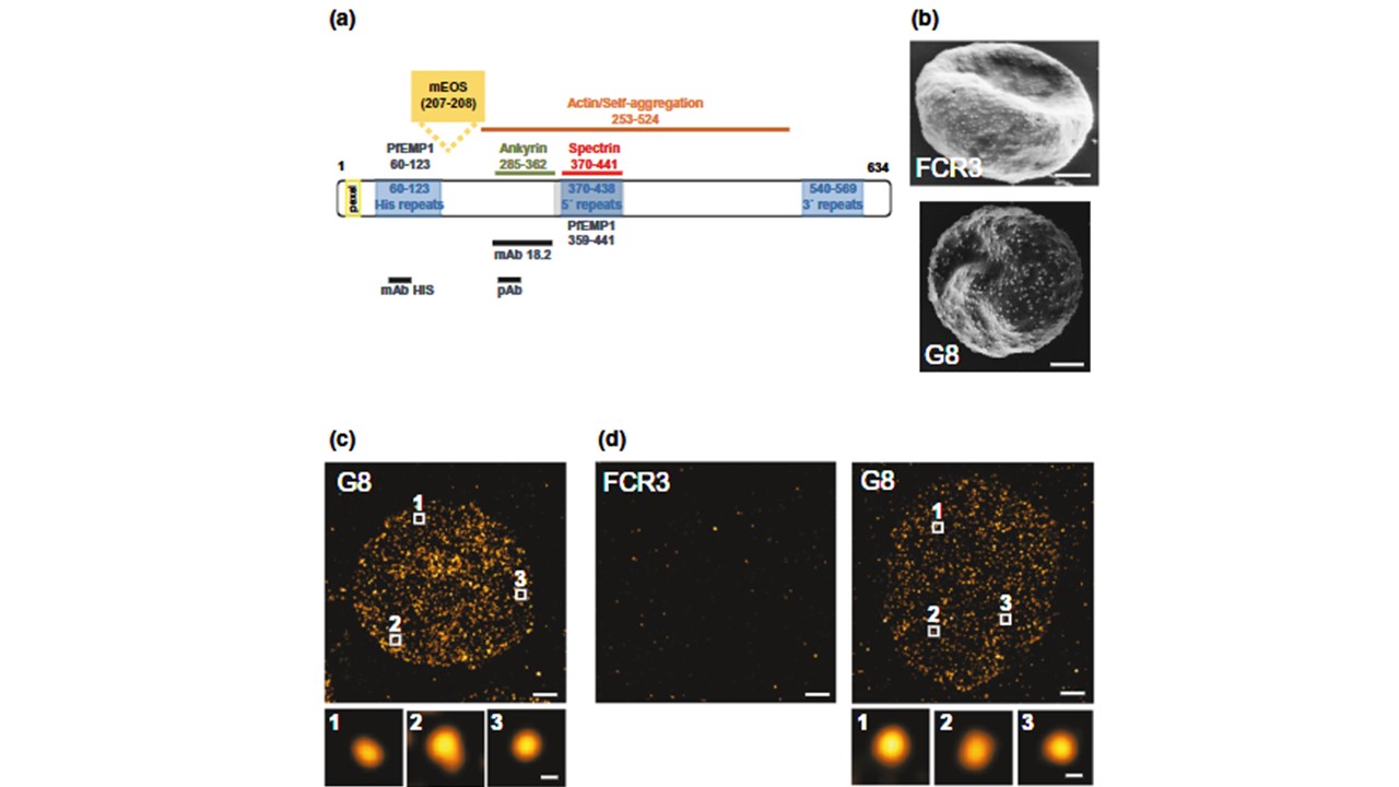
(a) Schematic illustration of KAHRP including structural features and interactions domains. The domains harboring epitopes for the monoclonal antibody mAB18.1, the peptide antiserum, and the monoclonal antihistidine antibody are indicated. mEOS2 was inserted between residues 207 and 208. (b) Representative scanning electron microscopic images of erythrocytes infected with the parental line FCR3 or the genetically engineered clone G8 expressing a genomically encoded KAHRP/mEOS2 fusion protein. Scale bar, 1 µm. The knob density per cell was determined and assessed by box plot analyses. Box plots show the individual data points, with the median (black horizontal line), the mean (red horizontal line) and the 25% and 75% quartile range being shown. Error bars indicate the 10th and 90th percentile. Statistical significance was assessed using the Mann–Whitney rank-sum test. (c) Representative dSTORM image of exposed membrane prepared from G8 trophozoites stained with the monoclonal anti-KAHRP antibodymAB18.2. Scale bar, 1 µm. Zoom-ins of boxed areas are shown below. Scale bar 50 nm. (d) Representative PALM images of FCR3 and the mutant G8 at the trophozoite stage. The numbered boxes refer to magnified fluorescence clusters presented in the lower panel column. Scale bars, 1 µm in the main panel and 50 nm in subpanels. (e) Number of KAHRP molecules per knob for G8 at the trophozoite stage, as determined by PALM. A total of 726 clusters from 15 cells were analyzed. A box plot analysis is overlaid over the individual data points Number of KAHRP molecules per Sanchez CP, Patra P, Chang SS, Karathanasis C, Hanebutte L, Kilian N, Cyrklaff M, Heilemann M, Schwarz US, Kudryashev M, Lanzer M. KAHRP dynamically relocalizes to remodeled actin junctions and associates with knob spirals in Plasmodium falciparum-infected erythrocytes. Mol Microbiol. 2022 117(2):274-292. 22. PMID: 34514656.
