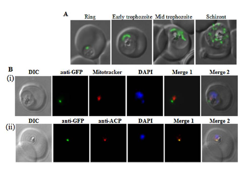
PfRRF1 localizes to the apicoplast.
A. Live cell images of PfRRF1leader–GFP
transfected parasites at different stages of the erythrocytic cycle. Signals indicative of apicoplast morphology are seen in the ring,
trophozoite and schizont stages.
B. Immunolocalization of PfRRF1. Panel (i) shows a differential interference contrast (DIC) image, nuclear DNA staining with DAPI, PfRRF1leader–GFP fluorescence, MitoTracker signal, and their overlap. Panel (ii) shows
colocalization of PfRRF1leader–GFP and the apicoplast marker ACP.
Gupta A, Mir SS, Jackson KE, Lim EE, Shah P, Sinha A, Siddiqi MI, Ralph SA, Habib S. Recycling factors for ribosome disassembly in the apicoplast and
mitochondrion of Plasmodium falciparum. Mol Microbiol. 2013 88(5):891-905.
PubMed Article: Recycling factors for ribosome disassembly in the apicoplast and mitochondrion of Plasmodium falciparum
Other associated proteins
| PFID | Formal Annotation |
|---|---|
| PF3D7_0208500 | acyl carrier protein |
