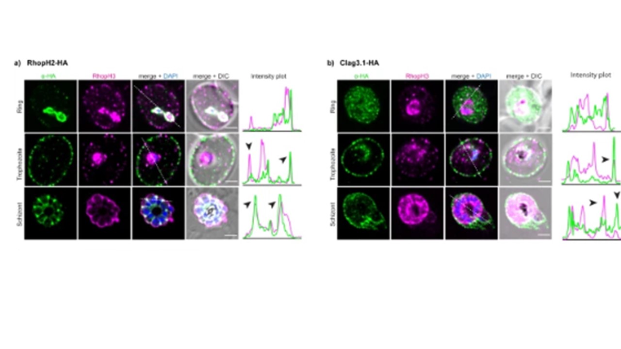
Super-resolution images showing colocalization of RhopH2-HA and RhopH3 during parasite development. Intensity plots along the white broken line show lack of colocalization at the ring stage, black arrows point at the points of colocalization on the erythrocyte surface and in the rhoptry. Scale bar 2 µm. b Super-resolution images showing colocalization of Clag3.1-HA with RhopH3 during parasite development. Intensity plots are shown along the white broken line that shows lack of colocalization at the ring stage, black arrows point at the points of colocalization on the erythrocyte surface and in the rhoptry. Scale bar 2 µm. c Heat map of the mass-spectrometry analysis from RhopH2-HA immunoprecipitation from developmental stages. d Immuno-blot (left panel) and quantification (right panel) of RhopH3 and Clag3 from RhopH2-HA immunoprecipitation of developmental stages. Error bars show SD from three biological replicates. Full blots shown. Pasternak M, Verhoef JMJ, Wong W, Triglia T, Mlodzianoski MJ, Geoghegan N, Evelyn C, Wardak AZ, Rogers K, Cowman AF. RhopH2 and RhopH3 export enables assembly of the RhopH complex on P. falciparum-infected erythrocyte membranes. Commun Biol. 2022 5(1):333. PMID: 35393572;
Other associated proteins
| PFID | Formal Annotation |
|---|---|
| PF3D7_0905400 | high molecular weight rhoptry protein 3 |
| PF3D7_0929400 | high molecular weight rhoptry protein 2 |