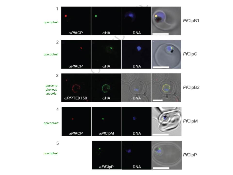
Localization of PfClpB1, PfClpC, PfClpB2, PfClpM, and PfClpP of P. falciparum was tested by immunofluorescent colocalization. In panels 1 and 2, PfClpB1-Strp-3×HA or PfClpC-Strp-3×HA parasites were probed with the apicoplast marker anti-ACP PFB0385w antisera (red), with anti-HA antisera (green), and with Hoescht 33342 to stain for DNA (blue). In panel 3, PfClpB2-Strp-3×HA parasites were probed with the parasitophorous vacuole marker anti-PTEX150 antisera (red), with anti-HA antisera (green), and with Hoescht 33342 (blue). In panels 4 and 5, wild type 3D7 strain was probed with anti-ACP antisera (red), with anti-PfClpM antisera or anti-ClpP antisera (green), and with Hoescht 33342 (blue). For all panels, the right most image represents the merged fluorescence images with the DIC or transmission image. The scale bars correspond to 5 mm.
El Bakkouri M, Pow A, Mulichak A, Cheung KL, Artz JD, Amani M, Fell S, de Koning T, Goodman CD, McFadden GI, Ortega J, Hui R, Houry WA. The Clp chaperones and proteases of the human malaria parasite Plasmodium falciparum. J Mol Biol. 2010 404(3):456-77
Other associated proteins
| PFID | Formal Annotation |
|---|---|
| PF3D7_0816600 | chaperone protein ClpB1 |
| PF3D7_1116800 | heat shock protein 101 chaperone protein ClpB2 |
| PF3D7_1406300 | glycerophosphodiester phosphodiesterase |
| PF3D7_1436300 | translocon component PTEX150 |
| PF3D7_API03600 | chaperone protein ClpM |
