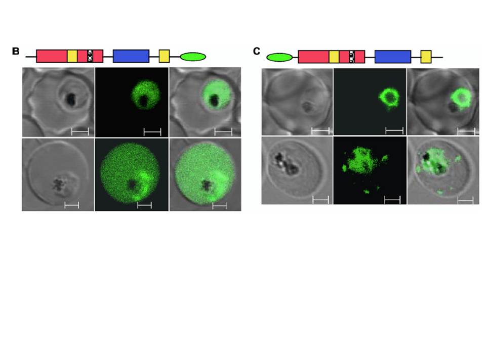
Live-cell imaging of GFP-tagged PfSec22 chimeras. (B) Live-cell imaging of parasites expressing the C-terminal GFP-tagged PfSec22 (PfSec22-GFP) showing diffuse localization of the protein throughout the parasite cytoplasm and occasionally in the host cell compartment. (C) Confocal micrographs showing localization of the N-terminal GFP-tagged protein (GFP-PfSec22) in earlytrophozoite (top row) and mid-trophozoite (bottom row) stage parasites. In addition to the ER-like profiles, GFP-PfSec22 associates with tubovesicular elements in the infected host cell. The micrographs (left to right) represent differential interference contrast, GFP fluorescence, and a merge of the two. Scale bars, 2 mm.
Ayong L, Raghavan A, Schneider TG, Taraschi TF, Fidock DA, Chakrabarti D. The Longin domain regulates the steady-state dynamics of Sec22 in P. falciparum. Eukaryot Cell. 2009 8(9):1330-40.