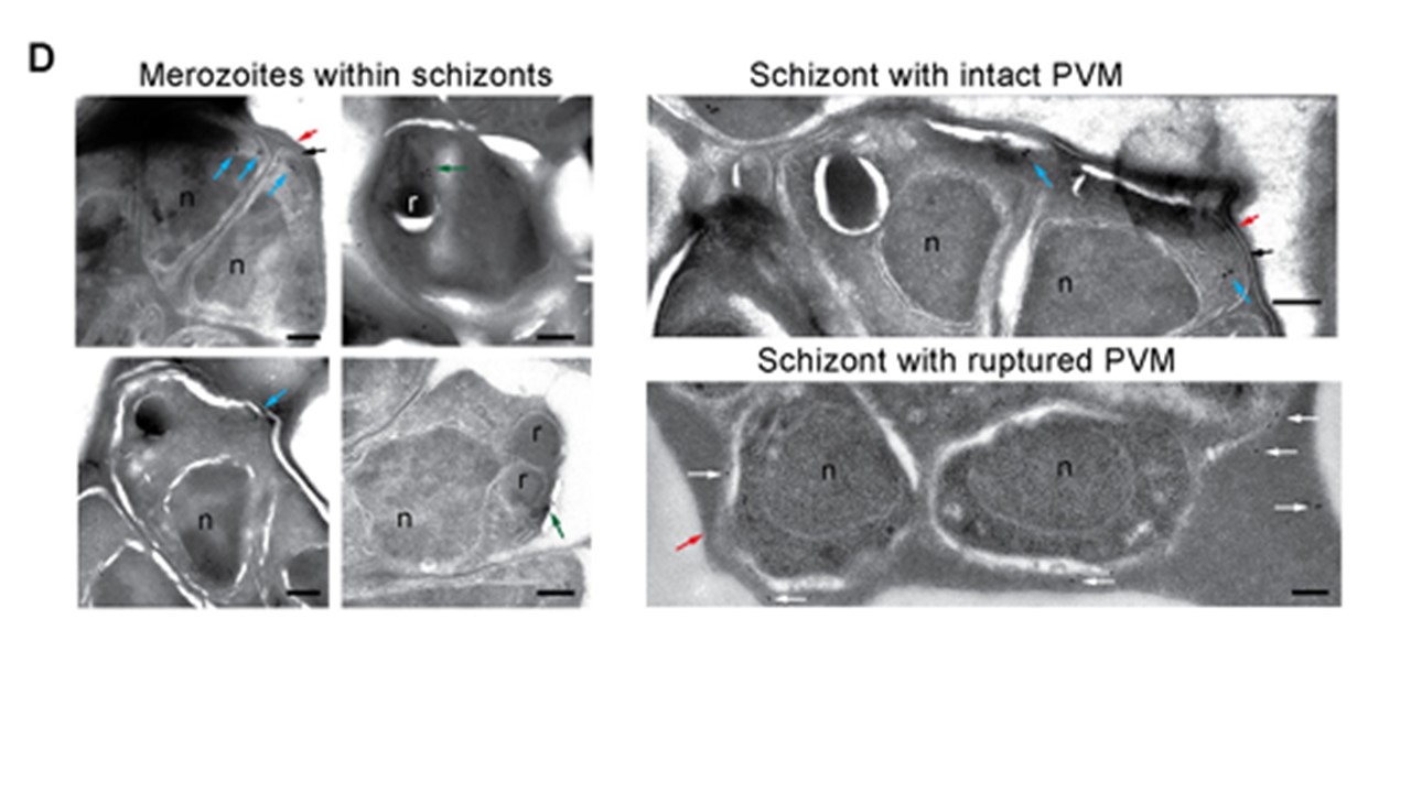
IEM section obtained from DPAP3-GFP parasites. Close-up images of individual intracellular merozoites on the left show immunogold staining of DPAP3-GFP in close proximity to the rhoptries (green arrows) and at the apical end of merozoites (blue arrows). Images on the right show representative sections of schizonts with an intact (black arrows) or rupture PVM. Staining of extracellular DPAP3-GFP(white arrows) was only observed in schizonts lacking a PVM. Rhoptries (r), nuclei (n), and the RBCM (red arrows) are indicated. Rabbit anti-GFP and colloidal gold-conjugated anti-rabbit antibodies were used. Bar graph = 200 nm. IEM images obtained on the 3D7 control line are shown in S3D Fig, and the uncropped IEM images for the DPAP3-GFP line in S3E . IEM section obtained from DPAP3-GFP parasites. Close-up images of individual intracellular merozoites on the left show immunogold staining of DPAP3-GFP in close proximity to the rhoptries (green arrows) and at the apical end of merozoites (blue arrows). Images on the right show representative sections of schizonts with an intact (black arrows) or rupture PVM. Staining of extracellular DPAP3-GFP (white arrows) was only observed in schizonts lacking a PVM. Rhoptries (r), nuclei (n), and the RBCM (red arrows) are indicated. Rabbit anti-GFP and colloidal gold-conjugated anti-rabbit antibodies were used. Bar graph = 200 nm. IEM images obtained on the 3D7 control line are shown in S3D Fig, and the uncropped IEM images for the DPAP3-GFP line in Lehmann C, Tan MSY, de Vries LE, Russo I, Sanchez MI, Goldberg DE, Deu E. Plasmodium falciparum dipeptidyl aminopeptidase 3 activity is important for efficient erythrocyte invasion by the malaria parasite. PLoS Pathog. 2018 14(5):e1007031. PMID: 29768491
