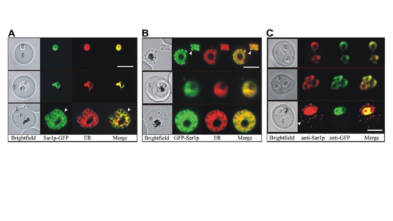
Subcellular location of the PfSar1p chimeras. (A and B) Co-labelling of the PfSar1p chimeras in transfected parasites with an endoplasmic reticulum (ER) probe. Live synchronous transfectants at different stages of the intraerythrocytic cycle were incubated with ER TrackerTM The first panel is a bright field image marked P (parasite) and E (erythrocyte). Bright puncta where the GFP is concentrated more than the ER Tracker are marked with arrows. (C) Dual immunofluorescence analysis of PfSar1p transfectants. Smears of erythrocytes infected with PfSar1p-GFP transfectants were probed with murine anti-GFP followed by anti-mouse IgG-Alexa Fluor 488 (green) and rabbit anti-PfSar1p, followed by anti-rabbit IgG-Alexa Fluor 568 (red). Both primary antibodies were used at 1:100. The first panel in each image set are bright field images. The arrow highlights structures in the infected erythrocyte cytoplasm. Bars denote 5 mm. Adisa A, Frankland S, Rug M, Jackson K, Maier AG, Walsh P, Lithgow T, Klonis N, Gilson PR, Cowman AF, Tilley L. Re-assessing the locations of components of the classical vesicle-mediated trafficking machinery in transfected Plasmodium falciparum. Int J Parasitol. 2007 37(10):1127-41. PMID: 17428488.
_,
