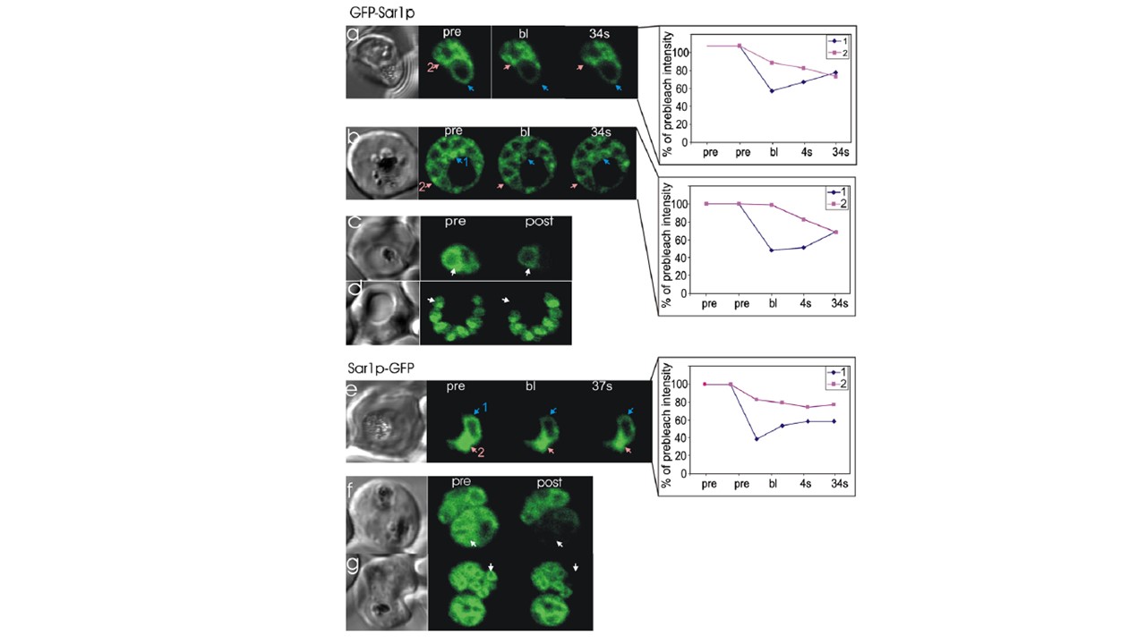
Photobleach analysis of GFP-tagged PfSar1p. Each series consists of a differential interference contrast image, and images showing pre-bleach
fluorescence (pre), fluorescence immediately after the bleach pulse (bl) and post-bleach images acquired after the times indicated. The position of the
bleach pulse is indicated by the number 1 and/or by an arrow. (a and b) GFP-PfSar1p: Application of a short laser pulse (200 ms) to trophozoite (a) or
schizont stage (b) parasites resulted in a decrease in fluorescence intensity in the bleach region (1) followed by rapid recovery at the expense of other
regions of the cell (2). Analysis of the fluorescence in the marked regions is presented in the plots at the right hand side. (c) Repeated bleaching of a region of a trophozoite stage parasite resulted in an even loss of fluorescence across the entire cell. (d) Bleaching of an individual merozoite in a very late stage schizont ablated the fluorescence from this parasite but did not affect other merozoites in the infected erythrocyte. (e) PfSar1p-GFP: Application of a short laser pulse (200 ms) to a trophozoite stage parasite resulted in a decrease in fluorescence intensity in the bleach region (1) followed by rapid recovery at the expense of other regions (2). (f) Repeated bleaching of a region of a doubly infected erythrocyte resulted in a relatively even loss of fluorescence across this cell but did not affect fluorescence in the other parasite. (g) Bleaching of an individual merozoite in a very late stage schizont ablated the fluorescence from this parasite but did not affect other merozoites. Adisa A, Frankland S, Rug M, Jackson K, Maier AG, Walsh P, Lithgow T, Klonis N, Gilson PR, Cowman AF, Tilley L. Re-assessing the locations of components of the classical vesicle-mediated trafficking machinery in transfected Plasmodium falciparum. Int J Parasitol. 2007 37(10):1127-41.. PMID: 17428488.
