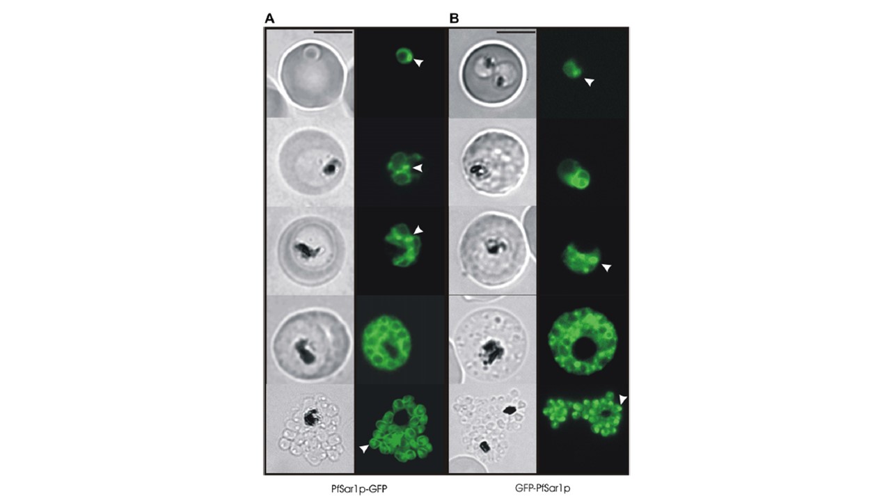
Expression of PfSar1p chimeras at different stages of the intraerythrocytic cycle of P. falciparum. The images represent differential interference contrast (DIC) micrographs and the GFP fluorescence signal for (A) PfSar1p-GFP and (B) GFP-PfSar1p. Ring and early trophozoite stage parasites (top rows) show a perinuclear ring of fluorescence in the parasite cytoplasm with bright puncta (arrows). Schizont stage parasites (fourth row) show rings of fluorescence around individual nuclei (see Supplementary Fig. S1). In burst schizonts (bottom row), a focus of bright GFP fluorescence is often observed at one end of the merozoite (arrows). Bar = 5 mm. Adisa A, Frankland S, Rug M, Jackson K, Maier AG, Walsh P, Lithgow T, Klonis N, Gilson PR, Cowman AF, Tilley L. Re-assessing the locations of components of the classical vesicle-mediated trafficking machinery in transfected Plasmodium falciparum. Int J Parasitol. 2007 37(10):1127-41. PMID: 17428488.
