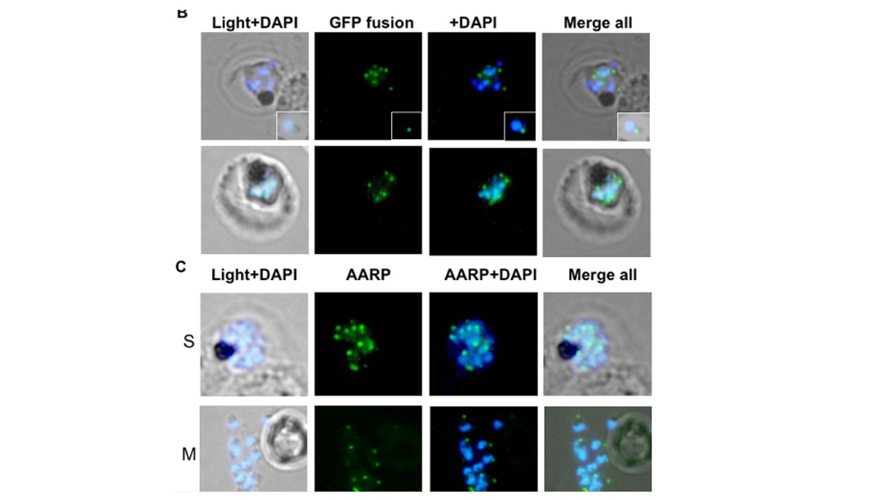
Localization of PfAARP to the apical end of the merozoites.
(B) Fluorescent microscopic images of transgenic parasites at schizont stages showing localization of PfAARP fused to a GFP reporter and expressed in an inducible system using schizont stage specific promoter. The parasite nuclei were stained with DAPI (blue). Enlarged images of selected individual free merozoite are shown in the insets. (C) Immuno-fluorescence assay to localize PfAARP in the schizont/merozoite stage parasites using anti-PfAARP (green) antibodies. The parasite nuclei were stained with DAPI (blue) and slides were visualized by fluorescence microscope. S, schizont and M, free merozoites. Wickramarachchi T, Devi YS, Mohmmed A, Chauhan VS. Identification and characterization of a novel Plasmodium falciparum merozoite apical protein involved in erythrocyte binding and invasion. PLoS One. 2008 3(3):e1732.