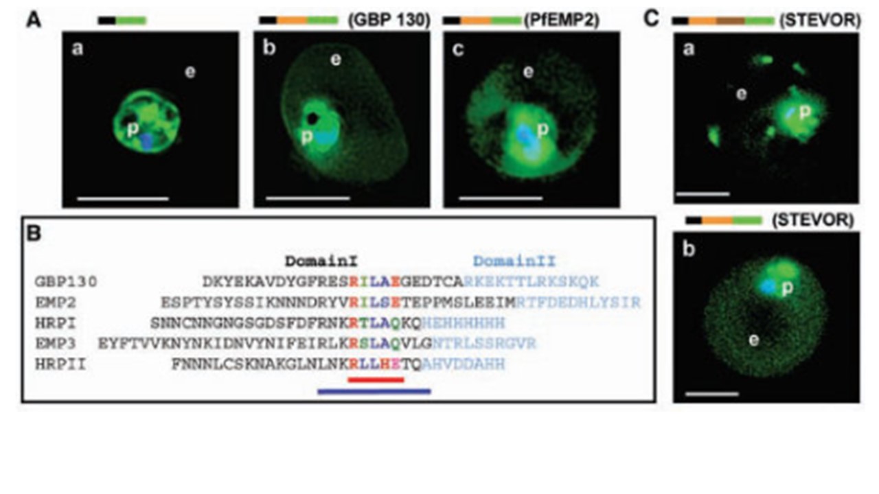
Identification of a conserved motif of 11 amino acids in VTSs of parasite proteins exported to the erythrocyte. (A) VTS of proteins PfGBP 130 and PfEMP2 target GFP to the erythrocyte. Projections (0-) of live cells expressing GFP chimeras of a SS alone (a), SSVTSGBP130 (b), and SSVTSEMP2 (c). (B) Alignment of MEME motifs in VTSs of indicated five exported soluble proteins. Red bar underlines 5–amino acid MEME motif 1. Blue bar underlines 11–amino acid MEME motif 2. (C) Projections (0-) of live cells expressing GFP chimeras of full-length STEVOR (a) and SSVTSSTEVOR (b). (A) and (C) were detected by digitized fluorescence microscopy. Parasite (p) nucleus is Hoechst stained (blue). Green, GFP; e, erythrocyte. Schematic above panels indicate constructs containing SS (black), VTS (orange) of the indicated proteins, and GFP (green). STEVOR sequences downstream of the VTS are indicated in brown in (C). Scale bars, 5 mm. Hiller NL, Bhattacharjee S, van Ooij C, Liolios K, Harrison T, Lopez-Estraño C, Haldar K. A host-targeting signal in virulence proteins reveals a secretome in malarial infection. Science. 2004 PMID: 15591203.
Ö¹s
Other associated proteins
| PFID | Formal Annotation |
|---|---|
| PF3D7_1016300 | glycophorin binding protein |