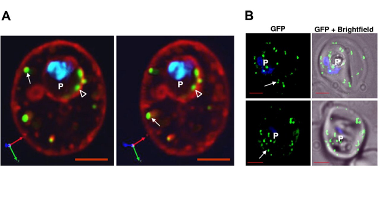
HT-dependent protein sorting into clefts may occur at parasite periphery and is not influenced by deletion of the C-terminal domain of PfSBP1. (A) Three-dimensional projections of a live infected erythrocyte expressing HT-GFPmembmyc and stained with TR-ceramide. Clefts at the periphery of the infected erythrocyte (arrows) as well as at or within the perimeter of the vacuolar parasite (empty arrowheads) are visible. (B) 0° projection of live infected
erythrocyte expressing HT-GFPmembmyc in 3D7 strains with parental (top) or chromosomal deletion of pfsbp1 (bottom), viewed under GFP optics and merged with bright field. Arrows indicate that the export of HTGFPmembmyc to cleft structures in parental 3D7 strain is not altered in parasite line with a C-terminal deletion in PfSBP1. Parasite (p) nucleus is stained with Hoechst 33342. Bar, 2 mm. Bhattacharjee S, van Ooij C, Balu B, Adams JH, Haldar K. Maurer's clefts of Plasmodium falciparum are secretory organelles that concentrate virulence protein reporters for delivery to the host erythrocyte. Blood. 2008 111(4):2418-26.