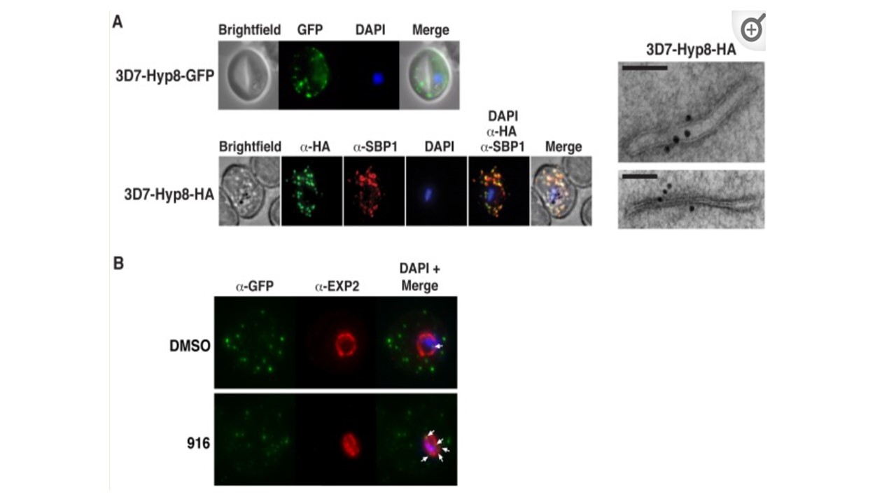
(A) (Top) Immunofluorescent micrographs show Hyp8-GFP is exported and localizes within puncta in the infected erythrocyte. (Middle) Hyp8-HA localizes within SBP1-containing MCs. (Right) Immunoelectron microscopy shows Hyp8-HA localization at MCs. Scale bar is 100 nm. (B) Maximum intensity projection micrographs showing export of Hyp8-GFP to MCs and secretion of EXP2 to the parasitophorous vacuole membrane following treatment with DMSO or 916 (50 mM). Puncta of nonexported GFP within the parasite and vacuole is shown (arrows). Sleebs BE, Lopaticki S, Marapana DS, O'Neill MT, Rajasekaran P, Gazdik M, Günther S, Whitehead LW, Lowes KN, Barfod L, Hviid L, Shaw PJ, Hodder AN, Smith BJ, Cowman AF, Boddey JA. Inhibition of Plasmepsin V activity demonstrates its essential role in protein export, PfEMP1 display, and survival of malaria parasites. PLoS Biol. 2014 12(7):e1001897. PMID: 24983235
Other associated proteins
| PFID | Formal Annotation |
|---|---|
| PF3D7_0113900 | Plasmodium exported protein (hyp8) |
| PF3D7_1471100 | exported protein 2 |