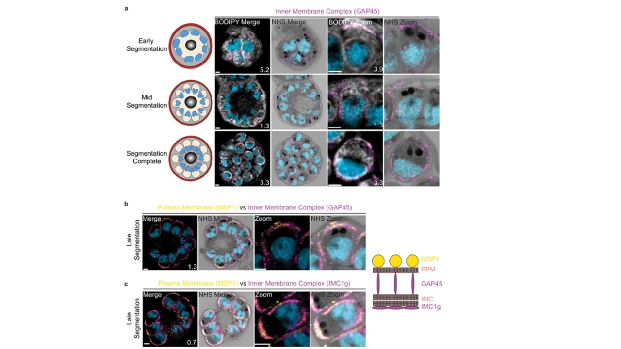
Inner membrane complex (IMC) progression through segmentation.
3D7 parasites were prepared by ultrastructural expansion microscopy (U-ExM), stained with N-hydroxysuccinimide (NHS) ester (grayscale), BODIPY TRc (white), SYTOX (cyan), and anti-GAP45 (PF3D7_0525800; IMC) (magenta) antibodies and imaged using Airyscan microscopy across segmentation. 3D7 parasites were stained with NHS ester, SYTOX, anti-MSP1 (parasite plasma membrane), and either anti-GAP45 (b) or anti-IMC1g (c) antibodies. GAP45 resides in the IMC luminal space, while IMC1g resides on the cytosolic side of the IMC. Neither GAP45 nor IMC1g could be reliably distinguished from MSP1. Images are maximum-intensity projections, number on image = Z-depth in µm of projection. Scale bars = 2 µm.
Anaguano D, Frölich S, Muralidharan V, Wilson DW, Dvorin J, Absalon S. Liffner B, Cepeda Diaz AK, Blauwkamp J, Anaguano D, Frolich S, Muralidharan V, Wilson DW, Dvorin JD, Absalon S. Atlas of Plasmodium falciparum intraerythrocytic development using expansion microscopy. Elife. 2023 Dec 18;12:RP88088. PMID: 38108809;y. bioRxiv [Preprint]. 2023 24:2023.03.22.533773. PMID: 36993606;
Other associated proteins
| PFID | Formal Annotation |
|---|---|
| PF3D7_1222700 | glideosome-associated protein 45 |