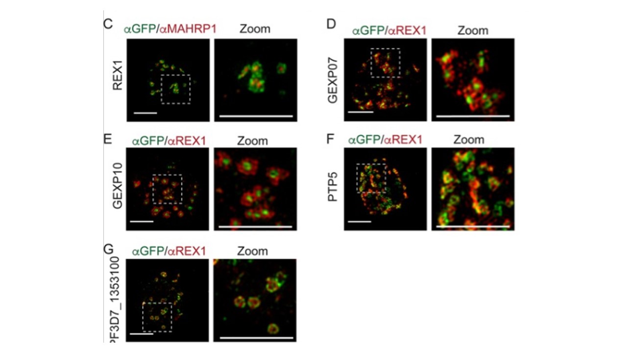
Maurer’s cleft proteins interact to form distinct protein clusters. (A and B) Protein interaction maps highlighting a putative PfEMP1 loading hub comprising GEXP07, MAHRP1, and GEXP10 (A) and a putative unloading hub comprising REX1, PTP5, SEMP1, and PF3D7_1353100 (B). Green nodes represent the GFP-tagged proteins. Double-thickness edges indicate reciprocal co-precipitation (see Fig. S3 for full-network maps). (C) 3D-SIM analysis of REX1-GFP-infected RBCs fixed and labeled with anti-GFP (green) and anti-MAHRP1 (red). (D to G) 3D-SIM analysis of transfectant-infected RBCs expressing GFP-tagged GEXP07, GEXP10, PTP5, and Pf3D7_1353100 that were fixed and labeled with anti-GFP (green) and anti-REX (red) antibodies. Maximum projections of Z-stacks are displayed. Scale bars = 3 μm, zoom scale bar = 3 μm. McHugh E, Carmo OMS, Blanch A, Looker O, Liu B, Tiash S, Andrew D, Batinovic S, Low AJY, Cho HJ, McMillan P, Tilley L, Dixon MWA. Role of Plasmodium falciparum Protein GEXP07 in Maurer's Cleft Morphology, Knob Architecture, and P. falciparum EMP1 Trafficking. mBio. 2020 11(2):e03320-19.
6
Other associated proteins
| PFID | Formal Annotation |
|---|---|
| PF3D7_0935900 | ring-exported protein 1 |
| PF3D7_1002100 | EMP1-trafficking protein |
| PF3D7_1353100 | Plasmodium exported protein, unknown function |
