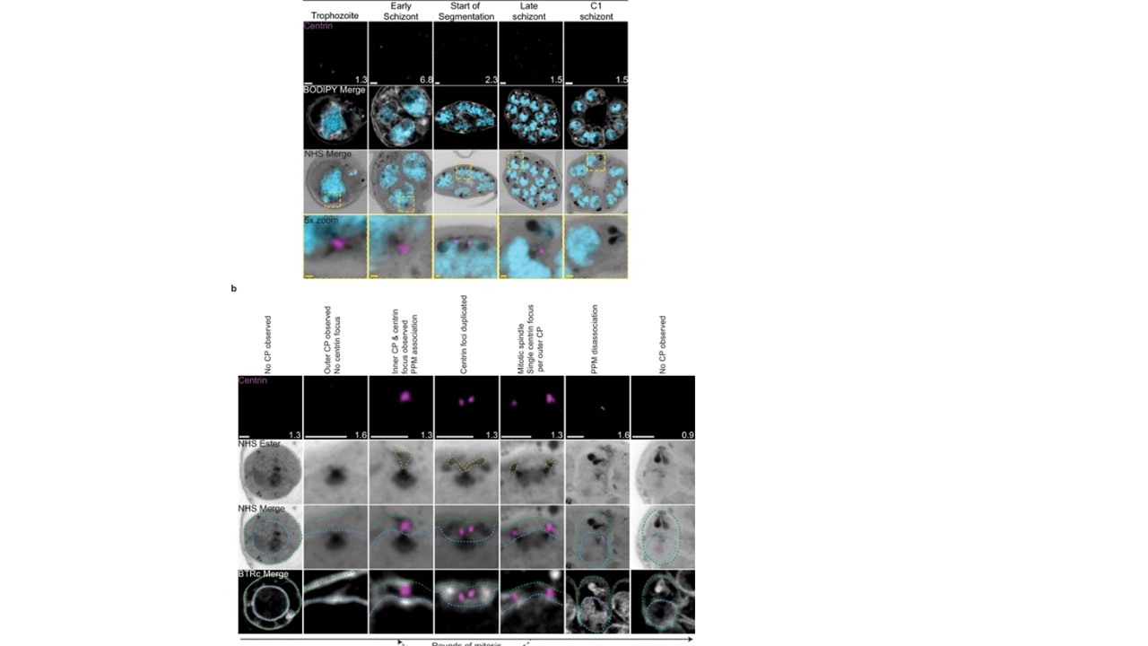
Centriolar plaque (CP) biogenesis and dynamics.
3D7 parasites were prepared by ultrastructural expansion microscopy (U-ExM), stained with N-hydroxysuccinimide (NHS) ester (grayscale), BODIPY TRc (white), SYTOX (cyan), and anti-centrin (outer CP; magenta) antibodies and imaged using Airyscan microscopy. (a) Images of whole parasites throughout asexual blood-stage development. (b) Whole parasite panel (left) followed by individual CP or CP pair zooms following our proposed timeline of events in CP biogenesis, dynamics, and disassembly. Yellow line = cytoplasmic extensions, blue line = nuclear envelope, green line = parasite plasma membrane. Images are maximum-intensity projections, number on image = Z-axis thickness of projection in µm. White scale bars = 2 µm, yellow scale bars = 500 nm.
Frölich S, Muralidharan V, Wilson DW, Dvorin J, Absalon S. Atlas of Plasmodium falciparum intraerythrocytic development using expansion microscopy. bioRxiv [Preprint]. 2023 9:2023.03.22.533773. Update in: Elife. 2023 Dec 18;12: PMID: 36993606
¿m