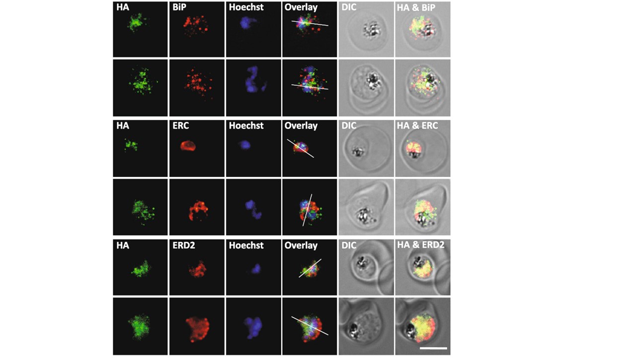
Colocalization of PfUT-HA with ER and Golgi markers in pfut mutants. Indirect immunofluorescence assay (IFA), using pfut mutants (at the trophozoite stage) and an anti-HA antiserum (mouse, 1:1000) together with the ER marker BiP (rabbit, 1:1000) or ERC (rabbit, 1:500), or with the Golgi marker ERD2 (rabbit, 1:500). Secondary antibodies were an anti-mouse Alexa Fluor 488 (green) and an antirabbit Alexa Fluor 546 (red). The nuclei were visualized with Hoechst (blue). Different channels, the differential interference contrast (DIC) image and two overlay images are shown for two representative examples each. PfUT is localized at the ER/Golgi complex in the mutants. A previous study has localized PfUT at the parasite’s ER/Golgi complex. Immunofluorescence assays, using the conditional knock-down mutants, revealed comparable results, with the HA-tagged PfUT partially co-localizing with the ER markers, BiP and ERC, and the Golgi marker, ERD2.
Jankowska-Döllken M, Sanchez CP, Cyrklaff M, Lanzer M. Overexpression of the HECT ubiquitin ligase PfUT prolongs the intraerythrocytic cycle and reduces invasion efficiency of Plasmodium falciparum. Sci Rep. 2019 Dec 4;9(1):18333.
Other associated proteins
| PFID | Formal Annotation |
|---|---|
| PF3D7_0917900 | PfHsp70-2 |
| PF3D7_1108600 | endoplasmic reticulum-resident calcium binding protein |
| PF3D7_1353600 | ER lumen protein retaining receptor |
