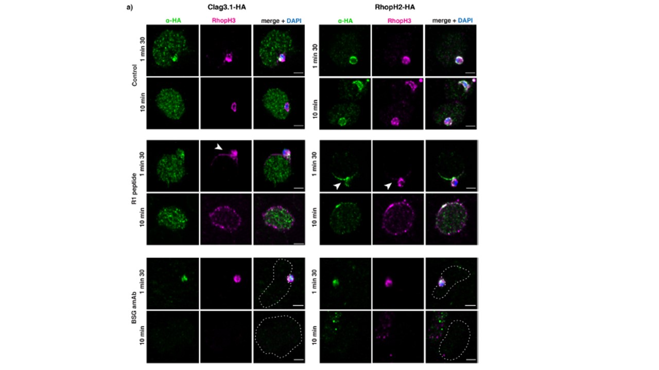
RhopH proteins have different cellular localizations during invasion. Super-resolution imaging of Clag3.1-HA, RhopH2-HA and RhopH3 (DMSO control). RhopH proteins in the presence of invasion inhibitors with DMSO control (top two panels), R1 peptide (middle two panels) and anti-BSG (bottom
two panels). Arrows point to membrane localization of RhopH2 and RhopH3. A dashed line outlines the erythrocyte. Scale bar 2 μm. Pasternak M, Verhoef JMJ, Wong W, Triglia T, Mlodzianoski MJ, Geoghegan N, Evelyn C, Wardak AZ, Rogers K, Cowman AF. RhopH2 and RhopH3 export enables assembly of the RhopH complex on P. falciparum-infected erythrocyte membranes. Commun Biol. 2022 5(1):333.
;
PubMed Article: RhopH2 and RhopH3 export enables assembly of the RhopH complex on P. falciparum-infected erythrocyte membranes
Other associated proteins
| PFID | Formal Annotation |
|---|---|
| PF3D7_0905400 | high molecular weight rhoptry protein 3 |
| PF3D7_0929400 | high molecular weight rhoptry protein 2 |
