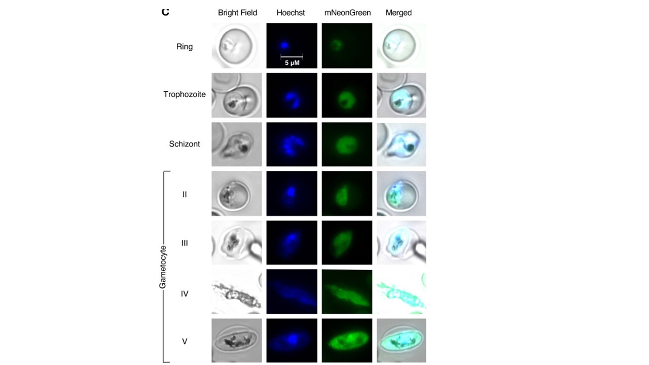
mNeonGreen@pare fluorescence varies between different asexual blood stages. (A) mNeonGreen@pare.2 parasites were sorbitol-synchronised and microscopy was used to confirm stages. Flow cytometry with MitoTracker DeepRed was used to enumerate parasites expressing green fluorescence in ring, trophozoite and schizont-staged cultures. FlowJo was used to gate single-cell, parasitised RBCs, quantify green fluorescence, and generate histograms. mNeonGreen@pare schizonts demonstrated the greatest fluorescence followed by trophozoites and then rings. (B) Stage-specific RNA-sequencing obtained from PlasmoDB shows that pfpare is expressed throughout the entire 48hr intraerythrocytic life cycle and expression peaks at late trophozoite and schizont stages (Chappell et al., 2020; Amos et al., 2021). (C) Fluorescence microscopy of synchronised mNeonGreen@pare parasites at asexual ring, trophozoite and schizont stages, and at gametocyte stages II-V (obtained through gametocytogenesis induction). Cultures were fixed, stained with Hoechst DNA stain, and visualised using bright-field and fluorescence microscopy at 1000x magnification. Hoshizaki J, Jagoe H, Lee MCS. Efficient generation of mNeonGreen Plasmodium falciparum reporter lines enables quantitative fitness analysis. Front Cell Infect Microbiol. 2022 12:981432. PMID: 36189342
aà"ma
