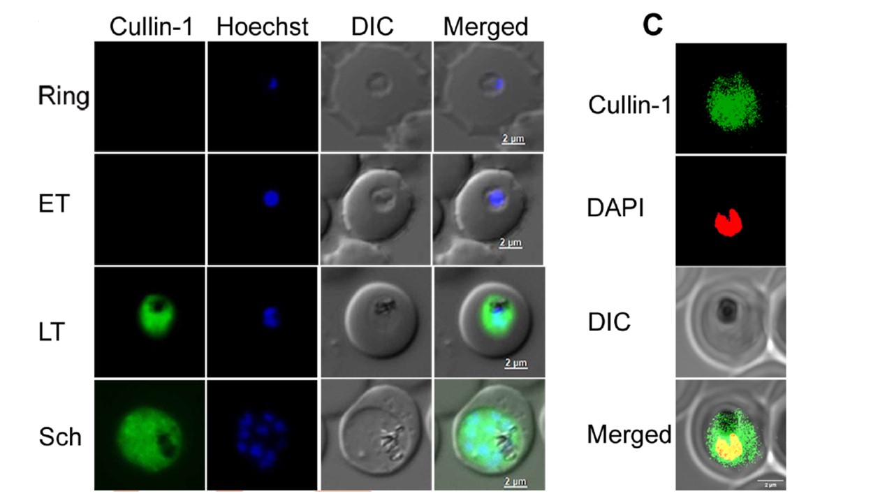
Expression and localization of PfCullin-1. A. The lysates of PfCul1GFPKI parasites corresponding to ring (R), early trophozoite (ET), late trophozoite (LT) and schizont (S) stages were assessed for expression of PfCullin-1/GFP and β-actin as a loading control by western blot using anti-GFP (ab-GFP) and mouse anti-β-actin-HRP (ab-Ac) antibodies, respectively. The full-length PfCullin-1/GFP protein (~128 kDa) is indicated by arrow head in the higher exposure panel (HE). B. The ring, early trophozoite (ET), late trophozoite (LT) and schizont (Sch) stages of PfCul1GFPKI parasites were assessed for localization of PfCullin-1/GFP by live-cell fluorescence microscopy. The panels show signal for PfCullin-1/GFP (Cullin-1), nuclear stain (Hoechst), bright field with RBC and parasite boundaries (DIC) and overlap of the three images (Merged). C. The confocal images of fixed PfCul1GFPKI trophozoite stage show colocalization of PfCullin-1/GFP with the nuclear stain DAPI (Pearson’s coefficient 0.59±0.10 from 27 images). The panels are as in B except that DAPI was used for nuclear staining. Rizvi Z, Reddy GS, Gorde SM, Pundir P, Das D, Sijwali PS. Plasmodium falciparum contains functional SCF and CRL4 ubiquitin E3 ligases, and CRL4 is critical for cell division and membrane integrity. PLoS Pathog. 2024 20(2):e1012045. PMID: 38416790;