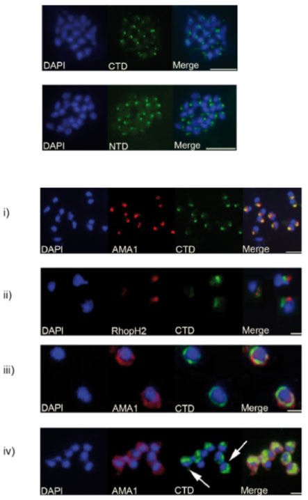
Analyses of the distribution of GAMA in asexual blood stage parasites by immunofluorescence. . Formaldehyde-fixed P. falciparum 3D7 schizonts were probed with mouse anti-CTD (C-terminal domains; top panel) or anti-NTD (N-terminal domains; lower panel) antisera, and an anti-mouse-FITC conjugate (green). Parasite nuclei were stained with DAPI (blue). Staining with both antisera resulted in a strong punctuate pattern, with a single point of fluorescence located away from the nucleus, and consistent with a location in the apical organelles. Scale bars represent 2 mm. (B) Formaldehyde-fixed free P. falciparum merozoites were probed with rabbit anti-CTD antiserum, and either mouse polyclonal anti-AMA1 (PF11_0344)-antibodies (i, iii and iv) or mouse mAb 61.3 (ii). Mouse antibodies were detected with an anti-mouse Alexa Fluor 594 conjugate (red), and rabbit antibodies with an anti-rabbit Alexa Fluor 488 conjugate (green). Parasite nuclei were stained with DAPI (blue). Scale bar in (i) 5 μm, in remainder 2 μm. In (i) and (ii) there is a clear apical localization of AMA1, RhopH2 (PFI1445w) and GAMA, with AMA1 and GAMA appearing to be co-localized; (iii) shows a circumferential localisation of both GAMA and AMA1 on the surface of free merozoites; (iv) shows a ‘cap’-like distribution of GAMA on the surface of free merozoites (arrowed), in contrast to the circumferential distribution of AMA1.
Hinds L, Green JL, Knuepfer E, Grainger M, Holder AA. A novel putative GPI-anchored micronemal antigen of Plasmodium falciparum that binds to erythrocytes. Eukaryot Cell. 2009 8:1869-1879. Copyright 2010.