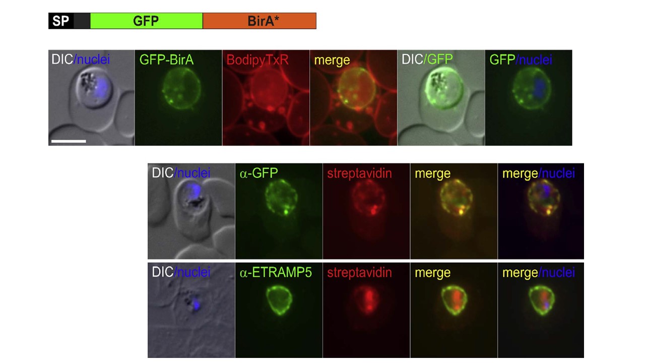
The SP-GFP-BirA* construct is located in the PV and is able to biotinylate proteins in this compartment. (A) Schematic view of the SP-GFP-BirA* construct. SP, Signal peptide; grey area: 16 amino acids left after the SP to serve as a linker before GFP. (B) Life cell images of a Bodipy-TX-ceramide stained parasite expressing the SP-GFP-BirA* construct. (D) Immune fluorescence assay (IFA) of SP-GFP-BirA* expressing parasites grown in the presence of biotin show co-localisation of biotinylated protein (detected using streptavidin) with GFPBirA* (upper panel) and ETRAMP5 (lower panel). (E) Western blot of saponin supernatant and pellet from parental (3D7) and SP-GFP-BirA* expressing parasites in the presence and absence of biotin. Biotinylation of proteins was visualized by streptavidin-HRP. Probable, self-biotinylated SP-GFP-BirA* is indicated with an arrowhead. Merge indicates overlay of green and red channels; size bar, 5 μm. Khosh-Naucke M, Becker J, Mesén-Ramírez P, Kiani P, Birnbaum J, Fröhlke U, Jonscher E, Schlüter H, Spielmann T. Identification of novel parasitophorous vacuole proteins in P. falciparum parasites using BioID. Int J Med Microbiol. 2017.
Other associated proteins
| PFID | Formal Annotation |
|---|---|
| PF3D7_0532100 | early transcribed membrane protein 5 |
