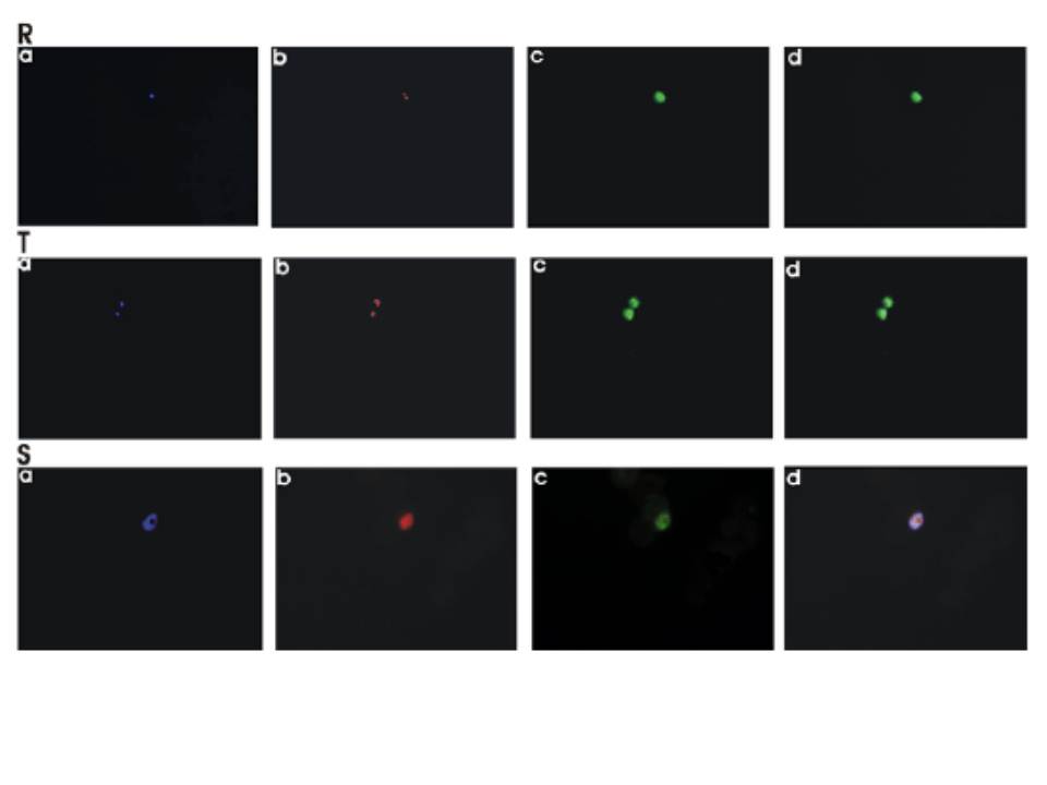
Immunofluorescence microscopy. Different fields were observed with excitation wavelengths of 350 nm (DAPI), 579 nm (MitoTracker Red CMXRos) and 489 nm (Cy2). Blue, red and green fluorescence indicate the location of (a) nucleus (b) mitochondria and (c) PfARP, in P. falciparum, respectively. (d) Merged picture of a, b and c pictures. Mononuclear (panel R), binuclear (panel T) and multinuclear (panel S) P. falciparum cells represent ring, trophozoite and schinzonts, respectively. PfARP expression predominated at ring and trophozoite stages. PfARP is a cytosolic protein.
Guha M, Choubey V, Maity P, Kumar S, Shrivastava K, Puri SK, Bandyopadhyay U. Overexpression, purification and localization of apoptosis related protein from Plasmodium falciparum. Protein Expr Purif. 2007 52:363-72.